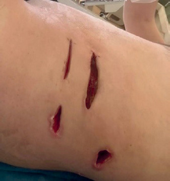
Necrotising fasciitis (NF) is a subset of aggressive skin and soft tissue infections that cause necrosis of the muscle fascia and subcutaneous tissue. NF is relatively rare, with an estimated annual incidence of NF of 0.5–10 cases per 100,000 people worldwide. This infection typically travels along the fascial plane, which has a poor blood supply, leaving the overlying tissues initially unaffected, potentially delaying diagnosis and surgical intervention [1, 2]. The infectious process can rapidly spread, causing infection of the fascia and peri-fascial planes, and leading to a secondary infection of the overlying and underlying skin, soft tissue, and muscle [3, 4]. Although it can occur in any region of the body, the abdominal wall, perineum, and extremities are the most common sites of infection. Involvement of the abdominal wall is usually a postoperative complication of abdominal surgery [1]. Elevated inflammatory biomarkers are standard features of NF patients. In one series, all instances of postoperative necrotising fasciitis occurred after contaminated or clean-contaminated surgery, mainly in patients with extensive faecal contamination of the abdominal cavity [1, 4–7].
A 35-year-old male was admitted to the hospital with the diagnosis of calculous cholecystitis. In accompanying examinations in the patient’s medical file, the abdominal ultrasound showed a gallbladder with thickened walls containing stones. Laboratory tests revealed WBC 9.68 × 10³ K/µl, haemoglobin 15.8 g/dl, total bilirubin 0.5 mg/dl, alanine aminotransferase (ALT) 51 U/l, and aspartate aminotransferase (AST) 20 U/l. The patient had no other accompanying diseases and did not report any history of alcohol, drug, or tobacco use. In these conditions, an uncomplicated laparoscopic cholecystectomy was performed. During the procedure, significant inflammation of the gallbladder was noted, along with numerous adhesions between the gallbladder and surrounding structures, particularly the duodenum. A closed suction drain was placed in the subhepatic space due to minor intraoperative oozing. The first 24-hour post-operative course following intervention was satisfactory. The next day, the patient experienced several episodes of fever, and a subcutaneous phlegmon (2–3 cm) was observed around the abdominal drain, which gradually evolved into cellulitis, rapidly spreading laterally and posteriorly along the external oblique muscle. Laboratory tests reveal WBC 19.6 K/µl, total bilirubin 1.3 mg/dl, amylase 564 U/l, and lipase 168 U/l. A consultation with an infectious disease specialist is held, and a computed tomography (CT) scan of the abdomen with oral and intravenous contrast is performed. The CT showed normal passage of the contrast through the stomach, duodenum, loops, and colon. However, the collection of tissue around the drain and along the external oblique muscle is considered necrotising fasciitis (infiltration of subcutaneous cellulo-fatty tissue and presence of gas bubbles). The abdominal drain is removed, and antibiotic therapy is added: imipenem and Tazocin IV (Figures 1, 2).
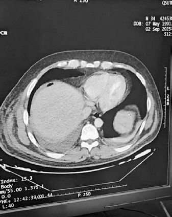
As soon as the results from the CT scan were obtained, we decided to perform drainage of the collection in several areas (three sites) with deep, communicating incisions in which drains were placed to help with wound treatment and drainage. In the following days, the antibiotic IV treatment continued, and the re-exploration of the wound was repeated (Figure 3).
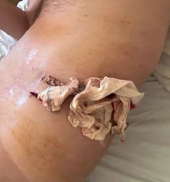
A repeat abdominal CT scan was performed, and despite a noticeable local improvement and the patient’s overall good condition, a decision was made to insert a new posterior drain due to a subcutaneous collection. A sample of the pus was sent for an antibiotic sensitivity test, which revealed the presence of Acinetobacter baumannii. In light of these results, in consultation with infectious disease specialists, the antibiotic regimen was modified to include imipenem, tigecycline, and clindamycin for further treatment.
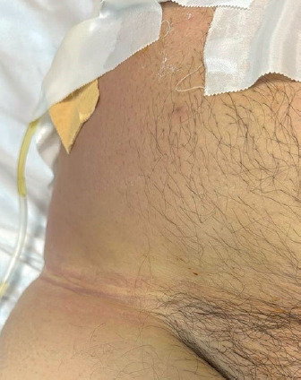
Antibiotic therapy was maintained postoperatively for 10 days, with local care strictly respecting the rules of asepsis. The evolution was favourable. The patient has been doing well for 1 month after the surgery (Figure 4).