
Crohn’s disease (CD) is characterised by chronic, transmural inflammatory response occurring in various segments of the digestive tract. The Crohn’s disease activity index (CDAI) is the primary clinical tool for determining the current disease severity. Despite progress in inflammatory bowel disease (IBD) pharmacotherapy, at least 20% of patients will experience a severe flare requiring hospitalisation. The management of the acute severe Crohn’s-associated colitis represents a significant medical challenge [1, 2]. The initial recommended management is intravenous steroids. Lack of significant clinical improvement within a week is an indication for therapy intensification. Prior to surgery, rescue therapy with biologics may be considered. In such cases, adalimumab and infliximab (IFX) may be effective; however, IFX seems to be the most effective due to weight-based dosing and generally higher doses used than those of adalimumab. In addition, IFX may be more efficient due to intravenous administration and slightly higher efficacy, and prompt action in mucosal healing [3–5]. Acute colectomy as rescue surgical treatment in CD is indicated in cases of megacolon and Crohn’s colitis resistant to previous therapeutic rescue options. Data about the effectiveness of rescue therapy in severe or fulminant CD flare is limited, and available results vary [6, 7]. The cumulative rate of colectomy in severe Crohn’s colitis seems to be higher than acute severe ulcerative colitis (UC). It results from high number of failures after conventional pharmacotherapy. Also, CD is considered as a disease with higher biological aggressiveness than UC due to its transmural involvement compared to the mucosal-only involvement in UC [8]. On the other hand, the rescue therapy for subjects with severe to fulminant CD exposed to biologics, including IFX, in the past remains a great challenge due to limited therapeutic possibilities. That is why there is an urgent need for the identification of new therapeutic options in those cases.
A 38-year-old woman with CD (diagnosed in 2002; Montreal classification: A1/L3/B1), as well as with psoriatic arthritis, Graves’ disease, fibromyalgia, vitiligo, osteopaenia, and micropituitary adenoma, was admitted to the Department of Digestive Tract Diseases in Norbert Barlicki Memorial University Hospital with severe hypogastric pain, fever with chills, arthralgia, and bloody diarrhoea (about 20 bowel movements per day) for several weeks with significant exacerbation in the last week before hospitalisation. So far, she had had two isotopic synovectomies and four arthroscopic synovectomies of both knee joints; however, she had not had any abdominal surgery. The family history was negative for IBD. At the onset of CD, the disease had an ileocolonic location. The upper part of the digestive tract had never been affected. The patient had a history of steroid dependence, and sulfasalazine and mesalamine intolerance since the CD diagnosis. Moreover, she had a history of IFX intolerance. It was the first biologic treatment for CD in this patient. After a dozen efficient doses, anaphylactic shock occurred at the next dose administration in 2022. In addition, complete inefficiency of azathioprine, 6-mercaptopurine, ustekinumab (drugs implemented for CD), golimumab, guselkumab, tofacitinib (drugs implemented for psoriatic arthritis), and methotrexate and adalimumab (drugs implemented for psoriatic arthritis) – the latter after initial effect for psoriatic arthritis – were shown in this patient. Furthermore, UPA at the dose of 15 mg was applied 12 months before the current hospitalisation due to exacerbation of psoriatic arthritis without satisfactory effect mainly for reduction of pain and oedema of joints. In addition, secukinumab, an anti-interleukin-17 antibody, was administered for psoriatic arthritis about 3 months before admission (four subcutaneous doses of 150 mg per week subsequently, 1 per month) with an increase of dose to 300 mg/month 8 weeks before the current hospitalization, which was accompanied by gradual exacerbation of CD symptoms, mainly bloody diarrhoea and abdominal pain.
At the current hospital admission, a mild normocytic anaemia level of haemoglobin of 10.7 g/dl (RR: 12.5–16.0 g/dl), C-reactive protein (CRP) 241 mg/l (RR: 0.0–5.0 mg/l), white blood cells 22.2 G/l (RR: 4.0–10.0 G/l), iron and vitamin B12 deficiency, and normal serum level of albumin 37.1 g/l (RR: 35–52 g/l) were seen. The faecal calprotectin (FCP) level was 665 µg/g. An intestinal ultrasound revealed colon wall thickening of about 8 mm in all segments. Abdominal and pelvic computed tomography (CT) at admission was negative for free fluid in the peritoneal cavity and intrabdominal masses, including abscesses. The acute severe flare of CD was diagnosed with a CDAI of 713 points. The patient did not respond to 5-day therapy with intravenous methylprednisolone (1 mg/kg/day). Ileocolonoscopy revealed erosion in the terminal ileum, deep ulcerations, loss of vascularity, spontaneous bleeding throughout the colon, and severe pancolitis (Simple Endoscopic Score for CD [SES-CD] – 35 points) (Figure 1 A). Histopathology showed cryptitis, crypt abscesses, and mixed inflammatory infiltrations primarily consisting of lymphoplasmocytes in the lamina propria. No cytomegalovirus (CMV) was detected in biopsy samples using immunohistochemical assay. Due to the lack of clinical response after intravenous steroids and former exposition to IFX, upadacitinib (UPA) at 45 mg/day orally was initiated as rescue therapy off-label. No rescue surgery was considered, due to the patient’s refusal. The choice of UPA was based on its mechanism of action, the literature data on its rescue potential, and the patient’s medical history. By the fifth day of UPA treatment, stool frequency had significantly decreased to a few per day without blood, and the CRP level was reduced to 95.5 mg/l. After notable clinical improvement, the patient was discharged, and careful clinical assessment was scheduled in a month.
Figure 1
Ileocolonoscopy images just before initiation of rescue therapy with upadacitinib (A), as well as after the first (B) and sixteenth (C) week of the treatment
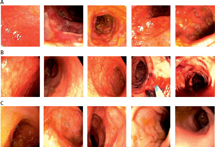
Unfortunately, after 5 days, the patient was again admitted to the same department with massive bleeding from the lower part of the digestive tract causing haemodynamic instability. Blood tests at admission revealed moderate normocytic anaemia and a level of haemoglobin of 9.4 g/dl, CRP level of 42 mg/l, mild hyponatraemia (Na 134 mmol/l), and hypoalbuminaemia (25.2 g/l). Due to haemodynamic instability and active bleeding, CT angiography of abdominal aorta with branches, renal, iliac, and femoral arteries was performed, which revealed extravasation of a contrast agent in the transverse colon (Figure 2). Ten units of packed red cells and four units of fresh frozen plasma were administered. Also, anti-haemorrhagics, including etamsylate and tranexamic acid, were applied. Interventional gastroscopy and ileocolonoscopy after preparation with enemas were performed. The gastroscopy showed no changes. In contrast, the ileocolonoscopy revealed a massive amount of blood and clots in the descending and sigmoid colon and rectum. After removal of blood and clots, two ulcers in the descending colon and one ulcer in the sigmoid colon, all with visible, protruding vessels with oozing bleeding, were detected, and haemostasis was reached with argon-plasma coagulation and metal clips. Also, ileocolonoscopy revealed normal terminal ileum and caecum, healing mucosa of the remaining parts of the colon with the presence of superficial ulcers in the transverse colon and deep ulcers in the descending and sigmoid colon, as well as the rectum. The decrease of the inflammatory changes (SES-CD – 21 points) compared to the ileocolonoscopy performed at the preceding admission (Figure 1 B) was seen. After the endoscopic examinations, bleeding was not observed, and the patient did not have diarrhoea. Intestinal ultrasonography was used as a non-invasive objective measure of treatment response, demonstrating a significant improvement with sigmoid and descending colon, and caecum wall width of 4 mm (Figure 3 A). Due to the significant endoscopic, sonographic, clinical (CDAI – 315 points), and biochemical response (CRP – 37.2 mg/l), the treatment with UPA was continued, and the patient was discharged with a stable level of haemoglobin and an oral diet.
Figure 2
Abdomen CT angiography scan presenting potential location of bleeding in transverse colon marked with red arrows
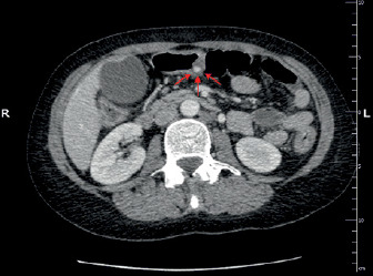
Figure 3
Intestinal ultrasonography images during the fourth (A) and sixteenth (B) week of the upadacitinib therapy
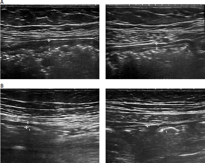
The UPA at the dose of 45 mg/day was continued for 12 weeks of the therapy, and subsequently the dose was reduced to maintenance of 30 mg orally. At the 16th week of therapy, the patient was in complete sonographic, biochemical, clinical, and endoscopic remission. CDAI scored 51 points. FCP level was < 30 µg/g, and CRP was 0.5 mg/l. The intestinal ultrasonography demonstrated the bowel wall thickening of 2–3 mm in sigmoid and descending colon and caecum (Figure 3 B). The ileocolonoscopy showed scars from healed ulcers and pseudopolyps in the transverse, descending, and sigmoid colon (SES-CD – 0 points) (Figure 1 C), and biopsies from the terminal ileum and all colonic segments showed mucosal healing and inactive inflammatory infiltrations. Currently, the patient remains clinically well without adverse effects of treatment, including infectious.
We present the case of a patient in whom a new approach to rescue therapy in acute severe CD was needed, and a new therapeutic option was applied off-label. Interestingly, acute severe Crohn’s-associated colitis flare was induced with the therapy with secukinumab used due to psoriatic arthritis. The likelihood that the acute severe CD flare was secukinumab-related was evaluated according to the Naranjo Adverse Drug Reaction probability scale and qualified as definite [9]. The inhibitors of IL-17 are commonly used for the treatment of psoriasis, psoriatic arthritis, and ankylosing spondylitis. The most recent data confirmed the association between the use of IL-17 blockers, including secukinumab, and the development of colitis, microscopic colitis, and exacerbation of IBD or IBD de novo. The detailed mechanisms of these paradoxical events are not fully understood. However, it seems that IL-17 may play a protective role for the gut epithelial barrier, and its blocking may lead to severe weakening of the gut epithelial barrier, intestinal inflammation induction, and the development of colonic infections. IL-17A-dependent regulation of occludin, which is a tight junction protein, limits gut permeability and maintains barrier integrity, while the absence of IL-17 signalling enhances inflammation [10]. Animal studies revealed that neutralisation of IL-17 by genetic ablation and antibody blocking led to severe weakening of the intestinal epithelial barrier and worsening of intestinal inflammation by enhancing gene expression of pro-inflammatory factors (IL-6, TNF-a, IFN-g) in colonic mucosa [11–13]. It was observed that IL-17 blocking may be efficient in other inflammatory disorders such as psoriasis, but it may cause exacerbation of CD [14]. However, our case is the first report associated with severe flare of IBD.
Therefore, all patients with IBD or IBD family history should be subjected to a careful, detailed clinical assessment, including FCP estimation, before administration of IL-17 inhibitors. In subjects with elevated FCP, complementary procedures should be introduced, such as colonoscopy and enterography [12]. Nevertheless, in the present case the patient had an ileocolonoscopy 1 week before the initiation of therapy with secukinumab, and this endoscopic examination showed no changes in the colon or terminal ileum. Therefore, the proposed precautions are not always sufficient, and secukinumab use should be avoided in IBD patients.
UPA is an oral, small molecule which is a selective inhibitor of Janus kinase 1 (JAK1), which plays a key role in the cytokine-mediated inflammatory response. UPA acts as an adenosine triphosphate-competitive JAK inhibitor, competing with ATP and blocking nucleotide binding to inhibit kinase activity and the phosphorylation of downstream effectors. It inhibits the formation of signal transducers and activators of transcription (STAT) dimers and consequently the expression of inflammation-related genes. UPA is registered, among others, for the treatment of moderate to severe UC and moderate to severe CD [15]. The most recent data show that UPA is characterised by high effectiveness in achieving and maintenance of clinical remission and endoscopic response in subjects with moderate to severe CD [16]. Currently, steroids and some biologics, primarily IFX, are the only therapeutic options for rescue management in severe to fulminant CD [5]. However, UPA has some features suggesting that it may be a candidate for rescue management of CD. The oral form of UPA is characterised by high bioavailability with rapid onset of action and short half-life. In addition, rapid absorption and quick clearance with low immunogenicity are other valuable features of this compound in rescue management [17, 18].
There are a few available studies assessing the potential utility of UPA in rescue therapy of IBD, mainly case series and studies. However, most of them were conducted in acute severe ulcerative colitis. The use of UPA in rescue management of CD, as in our case, represents novelty and off-label treatment. So far, there has only been one study assessing the efficacy of rescue therapy with UPA in acute severe colitis associated with CD. A multicentre retrospective cohort study by Clinton et al. evaluated the effectiveness of UPA in 12 patients with acute severe colitis in IBD, including three with CD. They were resistant to intravenous steroids and exposed to anti-TNF-a agents in the past. That is why the UPA at a daily dose of 45 mg was implemented as rescue therapy. In this study, the authors did not reveal detailed data about the efficacy of salvage treatment in this group of patients. Nevertheless, general clinical remission was achieved in 50% of all patients, and the colectomy rate at 90 days was only 25%. Interestingly, no adverse events, including serious one, were reported [1]. These results, along with our case report, are promising and point out the effectiveness and safety of rescue therapy with UPA. The rescue management of severe CD requires some modifications and a more individualised approach. The massive bleeding from the left colon that occurred a few days after the UPA initiation rescue therapy, in our opinion, was not related to this drug. It could result from the intensive colonic mucosal healing processes with extended inflammation and numerous ulcers. It should be also considered that bleeding in the course of IBD may be due to the protruding or oozing vessel, which should be additionally treated endoscopically.
In summary, we described a case demonstrating the effectiveness of UPA as rescue therapy in a patient with an acute severe CD flare following the failure of intravenous steroids and intolerance of IFX and other biologics in the past. The UPA treatment demonstrated rapid and sustainable clinical effects, achieving clinical, endoscopic, and histological remission for 4 months. In addition, this therapy seems to be safe and was not associated with adverse effects. Prospective studies, as well as comparative studies with available rescue therapeutic options like IFX, are necessary to assess the safety and efficacy of UPA in this indication. Nonetheless, UPA should be considered as a potential drug candidate for rescue management of IBD in the future.














