
INTRODUCTION
Chemerin, an adipokine encoded by the RARRES2 gene, has emerged as a mediator in various inflammatory disorders, as well as benign and malignant neoplasms. It also regulates numerous physiological and pathological processes through the chemerin/ChemR23 axis, which interacts with essential components of the immune response [1].
Serum chemerin levels have been associated with metabolic syndrome and its principal features, such as obesity, insulin resistance, and hypertension. Chemerin has also been shown to be markedly expressed in the dermis of early psoriatic lesions, where it contributes to the recruitment of plasmacytoid dendritic cells [2].
Psoriasis vulgaris is a chronic, immunemediated inflammatory skin disease characterized by keratinocyte hyperproliferation, angiogenesis, and immune dysregulation. Kong et al. demonstrated that chemerin enhances keratinocyte proliferation and stimulates the production of proinflammatory cytokines, thereby exacerbating psoriasis [3].
Psoriasis is further linked to vascular inflammation, oxidative stress, and dyslipidemia, which initiate and aggravate vascular injury and consequently increase the risk of cardiovascular disease (CVD) and metabolic syndrome. Elevated chemerin levels in psoriasis also facilitate monocyte–endothelial cell adhesion, leading to endothelial inflammation and atherosclerosis [4].
The early stages of cardiovascular disease often remain asymptomatic and are difficult to predict clinically. Therefore, serum biomarkers such as chemerin may serve as valuable screening markers to help identify patients at risk and guide further cardiac evaluation [5].
OBJECTIVE
The aim of this study was to assess the association between serum chemerin levels and subclinical cardiovascular changes in patients with psoriasis and to evaluate the potential role of chemerin as an early marker of cardiovascular involvement in this population.
MATERIAL AND METHODS
This study included 73 psoriatic patients, consecutively recruited, as group A from the dermatology outpatient clinic of Zagazig University Hospitals, and 73 healthy controls without psoriasis or any other inflammatory skin disease, recruited between April 2023 and September 2023. Participants aged over 60 years, smokers, and individuals with CVD, systemic diseases such as hypertension, hypo- or hyperthyroidism, diabetes mellitus, renal failure, dyslipidemia, or psoriatic arthritis were excluded. Recent use of biologic agents was also an exclusion criterion [6].
Written informed consent was obtained from all participants enrolled in the study. The study protocol was approved by the Institutional Review Board (IRB) of the Faculty of Medicine, Zagazig University.
Body mass index (BMI) was calculated for all participants, as weight divided by height squared (kg/m2). Waist circumference, systolic blood pressure (SBP), and diastolic blood pressure (DBP) were measured and recorded [7]. Severity of psoriasis in group A was assessed using the Psoriasis Area and Severity Index (PASI) score [8].
Laboratory assessment
Using an automated analyzer (Cobas 8000 platform, 720 module), total cholesterol, low-density lipoprotein cholesterol (LDL), high-density lipoprotein cholesterol (HDL), and triglycerides levels were measured in all participants.
Serum chemerin measurement
Serum chemerin levels were measured using human chemerin enzyme-linked immunosorbent assay (ELISA) kits provided by Sunred Biotechnology Co., Ltd. (China, catalogue no: 201-12-01436), according to manufacturer’s instructions.
Echocardiographic assessment
Echocardiographic images were obtained and recorded using standard techniques, including parasternal long-axis, parasternal short-axis, apical two-chamber, apical three-chamber, and apical four-chamber views. Left ventricular end-systolic (LVESD) and end-diastolic (LVEDD) diameters, left atrial diameter, left ventricular posterior wall thickness, and interventricular septal thickness were measured according to the American Society of Echocardiography guidelines. Left ventricular ejection fraction (LVEF), left ventricular end-systolic volume (LV-ESV), and left ventricular end-diastolic volume (LV-EDV) were measured using the modified Simpson method [9].
Apical four-chamber views with color-flow imaging were obtained to ensure optimal alignment of pulsed-wave (PW) Doppler with blood flow. Peak E-wave and A-wave velocities (cm/sec) and the mitral valve E/A ratio were measured. Tissue Doppler imaging (TDI) was then used to calculate mean septal and lateral basal e’ velocities and the mitral E/e’ ratio [10].
Left ventricular global longitudinal strain (LV-GLS) was analyzed by a specialized cardiologist who was blinded to study data, using a Vivid E9 commercial ultrasound scanner with phased- array transducers (M5S-D; frequency 2.0–4.0 MHz). Mean GLS was calculated by averaging the peak GLS values of apical two-chamber, apical three-chamber, and apical four-chamber images. Automatic endocardial margins were identified at end-systole. Manual adjustments were made when necessary to ensure accurate tracking and inclusion of LV wall thickness. A GLS value less negative than –18% was considered abnormal [9].
Statistical analysis
Data were analyzed using the Statistical Package for the Social Sciences software (SPSS, version 23.0). Comparisons between the psoriasis and control groups were performed using the independent samples ttest. Pearson’s and Spearman’s correlation coefficients were calculated to assess the relationships between serum chemerin levels and other variables. Multivariate linear regression analysis was conducted to identify independent predictors of GLS in patients with psoriasis. Receiver operating characteristic (ROC) curve analysis was applied to determine the optimal cutoff value for predicting subclinical systolic dysfunction.
RESULTS
Regarding demographic data, presented in Table 1, there were no statistically significant differences in age, sex, or smoking status between patients and controls. BMI was significantly higher in patients with psoriasis compared with controls. The Framingham Risk Score was applied to participants over 30 years of age to estimate the 10year risk of atherosclerotic cardiovascular disease (ASCVD), incorporating age, smoking status, systolic blood pressure, total cholesterol, and HDL cholesterol levels. The analysis revealed no significant differences between the two groups. Regarding laboratory findings, serum chemerin levels were significantly higher in the psoriasis group compared with controls. In addition, lipid parameters including total cholesterol, LDL, and triglycerides, were significantly elevated in the psoriasis group. Conversely, no statistically significant difference was observed in HDL levels between the two groups (table 1).
Table 1
Demographic and laboratory characteristics of the psoriasis and control groups
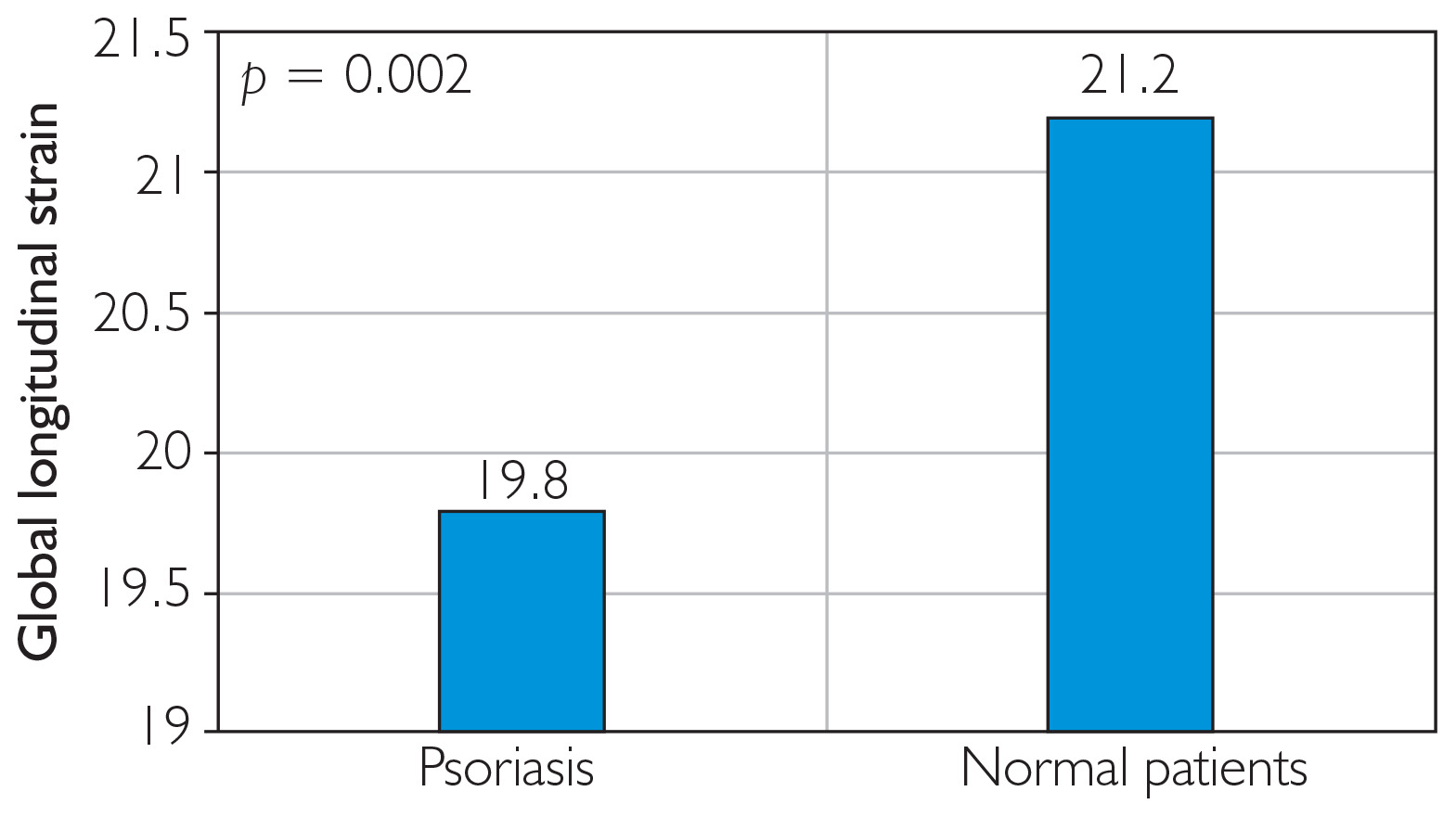
Regarding echocardiographic assessment of systolic function, LVEF was significantly lower in the psoriasis group (66% (51–78)) compared with the control group (70% (64–81)) (p < 0.05). Consistent with this finding, GLS, a marker of subclinical systolic dysfunction, was also significantly reduced in the psoriasis group (–19.8 Vs. –21.2, p < 0.05) (fig. 1). In contrast, E/e′, a marker of diastolic function, differed significantly between the two groups and indicated impaired diastolic function in patients with psoriasis (table 2).
Table 2
Echocardiographic parameters in the psoriasis and control groups
Figure 1
Comparison of global longitudinal strain between patients with psoriasis and healthy controls
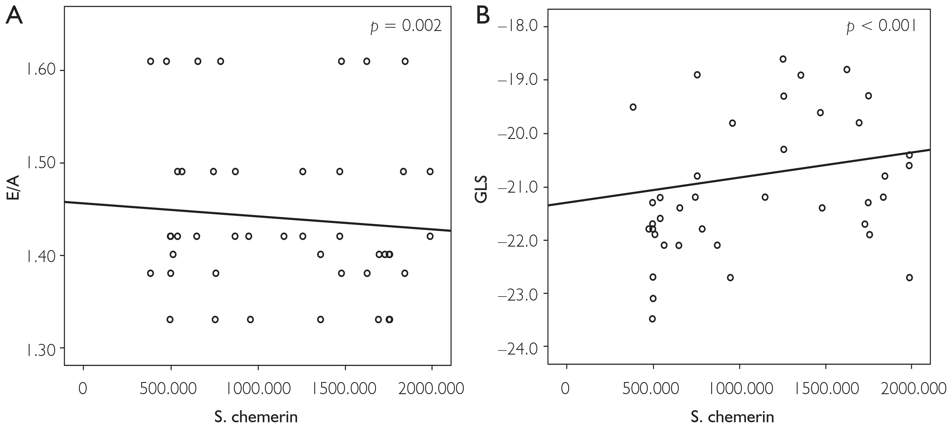
Correlation analysis revealed significant positive association between serum chemerin levels and age, BMI, PASI score, total cholesterol, triglycerides, LDL, E/e′, and GLS. Conversely, E/A ratio was significantly negatively correlated with chemerin levels (fig. 2). No significant correlation was observed between chemerin levels and LVEF (table 3).
Table 3
Correlations between serum chemerin levels and clinical, laboratory, and echocardiographic parameters
Figure 2
Correlations between serum chemerin levels and echocardiographic parameters. A – Significant negative correlation between serum chemerin levels and E/A ratio. B – Significant negative correlation between serum chemerin levels and global longitudinal strain (GLS)
Multivariate linear regression analysis showed that serum chemerin and BMI were independent predictors of GLS in patients with psoriasis (table 4). ROC curve analysis identified the optimal cut-off value of serum chemerin level for predicting subclinical systolic dysfunction based on GLS. Chemerin showed the best cut-off value for predicting subclinical systolic dysfunction at ≥ 2377.52, with a sensitivity of 87.5% and specificity of approximately 70.3 % (Supplementary table S1).
Table 4
Multivariable linear regression analysis with GLS as the dependent variable
DISCUSSION
Psoriasis is a chronic immunemediated skin disorder characterized by a complex inflammatory process aggravated by immune system dysregulation [10, 11]. These chronic inflammatory pathways initiate vascular endothelial dysfunction and platelet activation, which represent the earliest steps in the pathogenesis of atherosclerosis and subsequently increase the risk of CVD. Patients with psoriasis are up to 50% more likely to develop CVD, and this risk is positively correlated with the severity of skin lesions [12].
The association between psoriasis and CVD has been investigated in several studies, with both conditions found to share common risk factors. Harden et al. reported that CARD14 is predominantly expressed in epidermal keratinocytes, and its additional expression in aortic endothelial cells may represent a genetic risk factor in psoriatic patients [13]. Moreover, large prospective cohort studies have suggested that obesity and elevated BMI are independent risk factors for psoriasis [14]. Obesity is also associated with increased adipokine production, which contributes to endothelial damage, atherosclerosis, insulin resistance, and ultimately metabolic syndrome [15]. Another important link is smoking, a wellestablished risk factor for CVD, which has also been shown to exacerbate psoriasis severity [16].
At the cellular level, psoriasis and atherosclerosis share common inflammatory mediators, including tumor necrosis factor α (TNFα) and interleukin 1 (IL1). Platelets in psoriatic patients exhibit increased activation and cytokine release, particularly interleukin 1β (IL1β), a key cytokine involved in both psoriasis progression and the atherosclerotic process [17]. Immunemediated injury in psoriasis affects not only the skin but also the vasculature, leading to coronary microvascular dysfunction (CMD). Recent studies have demonstrated a positive correlation between psoriasis severity and CMD, identifying it as a poor prognostic factor even when clinically inapparent [18]. This subclinical involvement likely worsens cardiovascular outcomes and may contribute to increased mortality. Therefore, early detection of atherosclerosis in psoriatic patients may help reduce cardiovascular morbidity and mortality. Furthermore, recent evidence suggests that effective psoriasis treatment may lower cardiovascular risk, and some authors recommend incorporating psoriasis screening into preventive strategies [19, 20].
Chemerin is a novel adipokine that has recently attracted attention due to its role in metabolic, inflammatory, and carcinogenic processes [21]. It is a multifunctional chemoattractant protein implicated in the pathogenesis of both psoriasis and atherosclerosis. In psoriasis, chemerin activates the mitogen-activated protein kinase (MAPK) signaling pathway, recruits dendritic cells to sites of inflammation, enhances cytokine production, and promotes keratinocyte proliferation, thereby aggravating disease severity [3]. It has been shown to be more highly expressed in lesional skin compared to healthy tissue [22].
In the present study, serum chemerin levels were significantly higher in patients with psoriasis (p = 0.003) and positively correlated with PASI score (r = 0.339). This finding is consistent with the results of Aksu et al. [6]. While Coban et al. [23] confirmed the relationship between PASI score and chemerin, Gisondi et al. [24] did not, and Borsky et al. [5] reported no association.
Our study also demonstrated strong correlations between serum chemerin levels and age, BMI, total cholesterol, triglycerides, and LDL cholesterol. The association between chemerin and metabolic syndrome components has been well established in previous studies, which proposed that chemerin is a key regulator of adipocyte function, glucose metabolism, and other metabolic processes [25–27]. With respect to the cardiovascular system, chemerin has been implicated in the pathogenesis of hypertension, atherosclerosis, angiogenesis, and coronary artery disease [28]. Several studies have reported strong correlations between chemerin levels and CVD severity, although its role as a predictor of acute coronary syndrome remains unproven [29, 30].
In this study, LVEF and GLS were significantly lower in psoriasis patients compared with controls. These findings are in agreement with Pletikosic et al., who demonstrated reduced GLS in patients with moderate to severe psoriatic arthritis [31]. Conversely, Aksu et al. reported no significant difference in LVEF between psoriasis and control groups [6]. This discrepancy may be explained by differences in psoriasis severity among study populations, or by silent ischemia, in which LV systolic dysfunction remains clinically inapparent for years before symptoms manifest [32, 33]. These observations highlight the importance of GLS measurement in patients with psoriasis for the early detection of subclinical LV systolic dysfunction.
Regarding diastolic function, our findings revealed significant impairment in patients with psoriasis. This result is consistent with the studies of Aksu et al., Bülbül Sen et al., and Simsek et al., all of whom reported significant diastolic dysfunction in psoriasis compared with healthy controls [6, 34, 35]. These findings confirm the high susceptibility of patients with psoriasis to subclinical cardiac involvement.
Furthermore, our study demonstrated a significant positive correlation between serum chemerin levels and E/e′ as well as GLS. Multivariate linear regression analysis identified serum chemerin as an independent predictor of GLS in psoriasis patients. To our knowledge, this is the first study to assess the relationship between serum chemerin and GLS in psoriasis. This result is supported by Pletikosic et al., who reported strong associations between GLS, interleukin 17 (IL17), and adiponectin in psoriatic arthritis patients [31]. Proinflammatory cytokines, particularly IL17A, play a central role in the pathogenesis of inflammatory diseases, including psoriatic arthritis, and their effects on vascular and cardiac cells may contribute to the increased cardiovascular risk observed in these patients [36]. This may explain our findings regarding chemerin, a novel adipokine and multifunctional chemoattractant protein that activates MAPK signaling and is implicated in both psoriasis and atherosclerosis [22].
In this context, while Aksu et al. reported a significant positive correlation between serum chemerin and E/e′, they found no association with ejection fraction [6]. For this reason, our study focused on systolic function using GLS, which provides a more representative measure of subclinical systolic dysfunction.
This study had some limitations. The small sample size may restrict generalizability, and the cross-sectional design hinders causal inference. Potential confounders such as disease duration, systemic or biologic treatments and other confounding conditions could have influenced the results. Moreover, the single-center setting may limit external validity.
CONCLUSIONS
Patients with psoriasis are at increased risk of cardiovascular morbidity, which often remains clinically silent yet may significantly contribute to increased mortality. Early diagnosis of these changes remains challenging. The measurement of a simple and accessible blood biomarker such as chemerin may facilitate cardiovascular risk assessment in psoriatic patients, particularly in the presence of additional risk factors.












