
Introduction
Oesophageal and gastric cancers remain significant health concerns, with 1.48 million new cases and 1.10 million deaths reported worldwide in 2022 [1]. Esophagogastroduodenoscopy (EGD) is a common endoscopic procedure mainly used for detection and diagnosis of upper gastrointestinal neoplasm (UGN), which at an early stage may be resected endoscopically [2]. Endoscopic diagnosis of neoplasia at an early stage is especially demanded because early detection and subsequent minimally invasive resection lead to better survival rates [3]. The significance of quality in EGD has been emphasised in various endoscopy guidelines; in routine examination, it is estimated that 6–11.3 % of upper gastrointestinal cancers may be missed [4, 5]. A considerable number of these neoplasms originate from benign, precancerous conditions. One such example is “Correa’s cascade”, which occurs in the stomach, in which chronic atrophic gastritis progresses through intestinal metaplasia and dysplasia to invasive carcinoma [6]. Similarly, oesophageal adenocarcinoma develops from Barrett’s oesophagus and Barrett’s-related dysplasia [7]. For a high-quality search of precancerous and early neoplastic lesions, it is essential to thoroughly examine all three segments of the upper digestive tract and perform biopsies on any suspicious areas. Recently, new quality indicators have been validated, namely the composite detection rate (CDR) associated with the detection of UGN, and the endoscopist biopsy rate (EBR) related to missed upper gastrointestinal cancers [8, 9].
Coronavirus disease 2019 (COVID-19) presented a significant challenge with a high number of infections, a high mortality rate, and severe strain on the healthcare system [10].
Human-to-human transmission of SARS-CoV-2 occurs through respiratory droplets, aerosols, faeces, and contact with contaminated surroundings [11, 12]. Those characteristics make endoscopy a potential route for infection. In response to an elevated risk of virus transmission, endoscopic societies worldwide issued recommendations advising the use of personal protective equipment (PPE), including face masks with filters (N95, FFP2, or FFP3), face shields, and hair nets, in addition to gowns and gloves, as well as the temporary postponement of non-urgent examinations [13, 14]. Following these recommendations, upper endoscopy procedures were substantially reduced globally (on average by 82%) during the initial months of the pandemic [15]. Subsequent reports suggested that wearing PPE during the COVID-19 pandemic did not affect endoscopy quality indicators [16, 17]. Furthermore, a decline in endoscopy training has been noticed [18]. Finally, prolonged delays could result in a higher occurrence of mid- or long-term advanced cancer detection [19]. Our study aimed to determine the impact of the sanitary regime on the quality measures of EGD. Because the patients’ outcome is strictly related to the neoplastic disease stage, we investigated the influence of PPE on detecting neoplastic lesions, focusing on early ones.
Material and methods
Study design
This was a retrospective, multicentre study. Data from patients who underwent diagnostic EGD between January 2019 and December 2021 performed in three endoscopy units were analysed. According to the WHO, COVID-19 was declared a pandemic on 11 March 2020 [20], which was used as a reference date to compare the periods before and during the COVID-19 pandemic.
The research combined data gathered across three different centres: an endoscopy unit of a tertiary university hospital in Katowice (centre K), an endoscopy unit of a tertiary university hospital in Bialystok (centre B), and a private outpatient endoscopy clinic (centre P). The duration of the study was divided into 36-month periods.
Inclusion, exclusion criteria, and endoscopic procedure data
The inclusion criteria were as follows: age above 18 years, referral for diagnostic EGD.
The exclusion criteria were as follows: therapeutic procedures, incomplete examination due to intolerance, inadequate preparation, or upper GI obstruction, history of surgery (upper GI resections, except wedge gastrectomy), contraindications to biopsy (use of anticoagulant and antiplatelet drugs, portal hypertension, blood clotting disorders).
All endoscopists included in the study had completed endoscopic training and were gastroenterologists and internists. The procedures were performed on EVIS EXERA III and X1 systems (Olympus Europe, Hamburg, Germany) using GIF-EZ1500, GIF-1100, GIF-HQ190, GIF-H180J, GIF-H190, GIF-H180, and GIF-Q160Z endoscopes and Fujifilm EG-760R using the EP-6000 system (Fujifilm Europe, Düsseldorf, Germany).
Assessed parameters
The collected data included the following:
- Patient information: age, sex, indication for endoscopy;
- EGD data: inpatient or outpatient setting of the endoscopy, the endoscope type, sedation, name of the operator, if biopsy was obtained;
- Endoscopy report findings:
Oesophagus: cervical inlet patch, reflux esophagitis, Barrett’s oesophagus, Barrett’s oesophagus-related dysplasia, oesophageal lesion, oesophageal tumour, oesophageal subepithelial lesion,
Stomach: gastritis, atrophic gastritis, gastric intestinal metaplasia, gastric lesion, gastric peptic ulcer, gastric dysplastic lesion, gastric tumour, gastric subepithelial lesion,
Duodenum: duodenal bulb post-ulcer deformation, duodenal ulcer, duodenal polyp, duodenal tumour, duodenal subepithelial lesion.
The assessed quality measures were as follows:
- Composite detection rate (CDR) – the proportion of patients in whom at least one of the following lesions were detected: oesophageal inlet patch, gastric polyp, and post-ulcer duodenal bulb deformation.
- Endoscopist biopsy rate (EBR) – the proportion of examinations with at least one biopsy taken with the intention of histopathological examination.
- Upper gastrointestinal neoplasm (UGN) – the proportion of examinations that diagnosed a neoplastic lesion, namely dysplastic lesion, cancer, neuroendocrine neoplasm, stromal tumour, or lymphoma.
Dysplastic lesions were defined as low- and high-grade dysplasia focal lesions, described as 0-II (0-IIa, 0-IIb, 0-IIc) and 0-I (0-Ip, 0-Is) lesions according to Paris. Malignant neoplasms were defined as cancers, neuroendocrine neoplasms, stromal tumours, and lymphoma.
Statistical analysis
The quantitative measures of central tendency were calculated as mean ± standard deviation (SD) or median/interquartile range (IQR), taking into account the normality of distribution of the variable determined using the Kolmogorov-Smirnov test. For qualitative data, percentages and frequencies were calculated. The study was divided into 36 monthly periods. The correlation between monthly intervals and CDR, UGN, and EBR were calculated using the Kendall rank correlation coefficient. Comparisons of quantitative variables between two groups were performed using the paired samples t-test, Mann–Whitney test, or Wilcoxon signed-rank test for two groups determined by the normality of distribution and type of groups compared. The qualitative variables were compared using the χ2 test or Fisher’s exact test when appropriate. Univariate and multivariate logistic regression analyses were performed to assess the risk factors of detecting upper gastrointestinal dysplasia and neoplasms, and appropriate odds ratios (ORs) with 95% confidence intervals (95% CI) were calculated. In the multivariate analysis, we included variables that had a p-value lower than 0.1 in the univariate analysis. The data were analysed using IBM SPSS Statistics Version 29.0.1.0 (171) for MacBook.
Results
Study group characteristics
The study included a total of 12,322 diagnostic EGD procedures performed by 32 endoscopists (Figure 1). The median age of patients was 58 years (IQR = 24), and 56.1% of the patients were female, with 77.6% undergoing outpatient procedures. Out of all analysed cases, 40.9% were performed before the pandemic and 59.1% during the pandemic period. Demographic, endoscopic, and histopathological data are summarised in Table I.

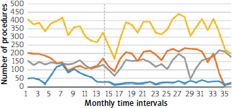
Table I
Demographic, endoscopic, and histopathological data before and during the COVID-19 pandemic
[i] COVID-19 – Coronavirus disease 2019. aAccording to European Society of Gastrointestinal Endoscopy (ESGE) Guideline. Weusten BLAM, Bisschops R, Dinis-Ribeiro M, et al. Diagnosis and management of Barrett’s oesophagus: European Society of Gastrointestinal Endoscopy (ESGE) Guideline. Endoscopy 2023; 55: 1124-46.
Endoscopic findings
During the investigated period 493 UGNs were found, 155 (3.1%) before and 338 (4.6%) during pandemic (p < 0.001). Of these, 288 patients were identified with dysplasia, 79 cases before and 209 during the pandemic (1.6% vs. 2.9% of diagnoses, respectively, p < 0.001). Malignant neoplasms were found in 213 EGDs, 78 before and 135 during the pandemic (1.6% vs. 1.9% of all EGDs, respectively, p = 0.204). Of the total, 8 patients were diagnosed with both dysplasia and malignant neoplasm. Two patients were simultaneously diagnosed with two different histological types of malignant neoplasms. Among all malignant neoplasms, there were 61 oesophageal tumours (30 squamous cell carcinomas, 22 adenocarcinomas, 1 anaplastic carcinoma, 1 lymphoma, 1 neuroendocrine tumour, 6 others), 143 gastric tumours (93 adenocarcinomas, 25 neuroendocrine tumours, 6 lymphomas, 4 squamous cell carcinomas, 3 anaplastic carcinomas, 3 undifferentiated carcinomas, 3 gastrointestinal stromal tumours, 6 others), and 11 duodenal tumours (7 adenocarcinomas, 3 neuroendocrine tumours, 1 lymphoma).
While Barrett’s oesophagus was diagnosed in 356 cases with no statistically significant difference before and during the pandemic identified (2.8% and 2.9%, respectively, p = 0.787) an increase of atrophic gastritis was observed (1527 patients; 10.6% vs. 13.6% before and during the pandemic, respectively, p < 0.001). The data are summarised in Table I.
Quality indicators before and during the COVID-19 pandemic
Within the analysed timeframes, increased UGN, CDR, and EBR were observed (Table II; Figures 2–4). During the COVID pandemic increased detection of both malignant tumours and dysplastic lesions was observed. The number of UGNs increased during the COVID pandemic with an OR of 1.53 (95% CI: 1.26–1.86) and a greater increase of dysplastic lesions (OR = 1.85; 95% CI: 1.43–2.41) rather than advanced malignant neoplasms (OR = 1.2, 95% CI: 0.91–1.59) – Table III.
Table II
Quantity of upper gastrointestinal dysplasia, neoplasm, and gastroscopy quality metrics before and during the COVID-19 pandemic among centres

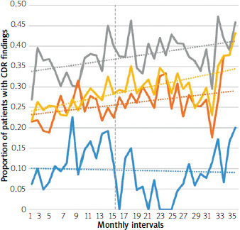

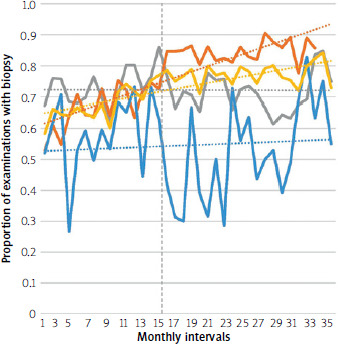

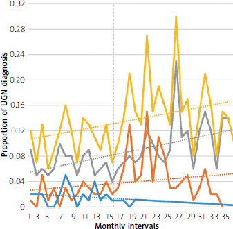
Table III
Comparison of EBR, CDR, UGN, dysplasia, and malignant neoplasm diagnosis in the upper digestive tract before and after the COVID-19 pandemic: univariate regression analysis
| Parameter | Odds ratio | 95% confidence interval | P-value |
|---|---|---|---|
| EBR | 1.69 | 1.56–1.83 | < 0.001 |
| CDR | 1.30 | 1.20–1.41 | < 0.001 |
| UGN | 1.53 | 1.26–1.86 | < 0.001 |
| Dysplasia | 1.85 | 1.43–2.41 | < 0.001 |
| Malignant neoplasm | 1.2 | 0.91–1.59 | 0.20 |
Risk factors for cancerous and precancerous conditions
In the multivariate regression analysis, the identified factors related to neoplasm detection were age, male sex, presence of alarm symptoms, EGD performed during the COVID-19 pandemic, and endoscopy performed in centre K (Table IV). When taking into consideration only dysplastic lesion detection, the risk factors were age, male sex, alarm symptoms, EGD performed during the COVID-19 pandemic, and endoscopy performed in centres K and P. The data are summarised in Table V.
Table IV
Logistic regression analysis of upper gastrointestinal neoplasm detection risk factors
Table V
Logistic regression analysis of upper gastrointestinal dysplasia detection risk factors
Comparison of endoscopy centres
Due to the association between endoscopic centres and neoplasia detection, as a secondary objective, a subgroup analysis was performed. The UGN detection rate was significantly higher in centre K than in centres P and B (6.2% vs. 2.2% vs. 2.5%; p < 0.001) (Figure 4). The upper gastrointestinal neoplasm detection rates before and during COVID-19 were found to have statistically significant differences when comparing data from all centres (3.1%. vs. 4.6%, < 0.001) (Table II). An increase in UGN was observed in centres K and P along with an increase in the detection of dysplastic lesions. There were no significant differences in the detection of malignant neoplasms (Table II).
The highest CDR was observed in centre K (37.7%) and the lowest (10.7%) in centre B (p < 0.001). In centre P the biopsy rate reached 77.5%, compared to centres K and B, where the EBR was 72.4% and 56.8%, respectively (Table II; Figures 2, 3). Also, EBR increased during the COVID pandemic in centres P and B, and CDR increased in centres K and P.
Discussion
EGD is crucial in the diagnosis and treatment of gastrointestinal diseases; however, as a procedure it carries a high risk of respiratory infectious disease transmission. To prevent infection, after the beginning of the pandemic, many medical facilities decreased the number of procedures compared to the same period in 2019 [21, 22]. The COVID-19 pandemic presented a formidable challenge for cancer patients because pursuing treatment exposed them to the risk of infection, while refraining from treatment significantly raised the risk of mortality [23].
We present data from three high-volume centres of different settings. The strength of our study is the comprehensive analysis of 12,322 EGDs from all three institutions. Our findings demonstrate that regardless of the operator’s use of PPE during the COVID-19 pandemic, the quality of EGD procedures was not compromised. This finding may be transferred to patients with similar infectious diseases with an indication for endoscopy or future infectious diseases of worldwide spread. In the last few years, quality metrics of EGD have been proposed. We opted to use the EBR and CDR because both can be calculated based on analysis of endoscopy reports. The improvement of quality parameters, namely CDR and EBR (Table II; Figures 2, 3), can be explained by the increased caution and awareness of endoscopists due to the deferment of non-urgent procedures and a focus on selected cases. The increased incidence of UGN during the pandemic (Table 2; Figure 4) corresponds with findings from other research studies [24, 25]. It is potentially caused by unconsciously increased the operator’s attention and awareness of disruptions during examinations due to PPE use. Also, it may be related to strict qualification for EGD. At the onset of the pandemic, the World Endoscopy Organisation issued recommendations on infection prevention in digestive endoscopy, based on experiences from China. For patients with mild symptoms and routine screening, EGD was recommended once the coronavirus outbreak had resolved [26, 27]. On the other hand, in centres where UGN was higher, an improvement in CDR was observed. It is important to note that we observed a higher rate of dysplasia detection, despite the lower median age during the COVID-19 pandemic. The only centre where neoplasia detection was not significantly increased was the centre in which the number of patients was lowest. This might be explained by the shift of endoscopy units working habits, focusing on emergency procedures, and the consequent selection bias, which was a commonly seen issue [28]. On the other hand, in the same centre CDR did not increase, so it might be speculated that the quality of examinations did not improve as much as in the other two centres. The consistent detection rates of Barrett’s oesophagus before and during the pandemic may be attributed to clearly recognisable endoscopic characteristics, which are less influenced by using PPE. A comparison between centres was conducted as a secondary objective to assess the impact of institutional differences on outcomes. Centre affiliation was included in the multivariate analyses to control for this variability. To address differences in treatment regimens during the COVID-19 pandemic, we presented both overall and centre-specific data, allowing for a more comprehensive evaluation of centre-related factors.
Despite other well-established risk factors of upper gastrointestinal neoplasia diagnosis such as age and male sex, we found out that use of high-definition endoscopes was neither related with higher capability to detect neoplastic lesions nor dysplastic (early) lesions. This is in line with previous reports, in which “western” high-definition endoscopes did not improve the detection of upper gastrointestinal lesions [8, 29].
It seems to be conflicting with available data showing that the use of high-definition equipment was found to be significantly better in the diagnosis of Barrett’s oesophagus-related dysplasia and early gastric lesions than standard-definition endoscopes [30, 31]. We put forward a hypothesis that equipment is less important than the operator’s capability to detect and correctly assess neoplasia in the upper digestive tract.
During the pandemic, there was an increase in the number of outpatient procedures, which may be related to a reduction in hospital admissions. There was also a significant reduction of performed diagnostic endoscopies visible from March to April 2020, compared to March–April 2019, 2021 (Figure 1). Nevertheless, the number of restricted procedures was lower than reported in most other countries [32, 33].
Our study has several limitations. First, examination time, another indicator of quality for EGD [34], was not considered in our analysis due to the retrospective character of the study. Second, in centre P, a private outpatient endoscopy clinic, the lowest incidence of UGN was detected, which could be potentially attributed to selection bias. Alternatively, the highest UGN detection rate in the tertiary centre may be attributed to patient preselection, leading to a higher proportion of patients with precancerous conditions and an increased risk of developing neoplastic lesions.
Third, not all procedural data, specifically sedation methods and outpatient/inpatient settings, were included in the multivariate analysis due to considerable heterogeneity between endoscopy centres. One centre used both conscious sedation and propofol anaesthesia, while the other employed only a single sedation method. Additionally, one centre conducted all procedures exclusively in an outpatient setting, whereas the other included single-day hospital admissions. This variability prevented the inclusion of these factors in the multivariate regression analysis.
Conclusions
We demonstrated that EGD quality measures were not compromised by the changes in working habits and the use of PPE during the COVID-19 pandemic. Additionally, we found an increase in neoplastic lesion detection during the pandemic, particularly among dysplastic lesions, along with other quality measures.














