
Introduction
The pancreas is an organ that fulfils both endocrine and exocrine functions. These functions work together to facilitate the processes of digestion, absorption, and metabolism of foodstuffs [1].
Pancreatic exocrine insufficiency (PEI) is a condition characterised by a deficiency of pancreatic enzymes, which can result in impaired digestive processes [2–4]. It is hypothesised that PEI may be a prevalent condition among patients with diabetes, potentially resulting from primary loss of parenchymal function and/or secondary impairment of pancreatic exocrine function and insufficient pancreatic enzyme activity.
A diagnosis of pancreatic and exocrine function can be made with confidence based on the results of the FE-1 concentration test. It is crucial to recognise that these findings should be interpreted with consideration and sensitivity to the patient’s condition. A concentration above 200 µg/g of faeces is evidence of normal function without abnormality, while a result between 100 µg/g and 200 µg/g is indicative of mild exocrine pancreatic insufficiency. Conversely, a result below 100 µg/g indicates severe exocrine pancreatic insufficiency [5].
A limited number of studies, primarily comprising small patient groups, suggest that PEI affects approximately 51% (26–74%) of individuals with type 1 diabetes and 32% (28–36%) of those with type 2 diabetes [1–3, 5–9].
The clinical picture and morphological changes of the pancreas in PEI in patients with diabetes do not correspond to the symptoms of typical chronic pancreatitis. There is a clear distinction between the two conditions. It has been demonstrated that the volume of the pancreas is only moderately decreased in patients with type 2 diabetes in comparison to the control group [8, 9]. It is important to note, however, that this group of patients demonstrates a higher degree of interlobular fibrosis and lobular atrophy in comparison to the control group. It is proposed that this particular case should be considered as a separate case and given the new name: ‘diabetic exocrine pancreatopathy’. The term is also referred to as ‘diabetic exocrine pancreatopathy’ [10–12].
Aim
The primary outcome measure was the incidence of PEI in diabetic patients. The secondary outcome measure was the evaluation of additional risk factors for PEI.
Material and methods
The study was retrospective and was conducted in 5 medical centres in Poland: National Medical Institute of the National Medical Institute of the Ministry of the Interior and Administration in Warsaw, Department of Gastroenterology and Hepatology with Endoscopy Unit, Medical University of Lublin, Department of Gastrointestinal Diseases of the Medical University of Lodz, Department of Gastroenterology, Allergology, and Paediatrics ICZMP of the Polish Mother’s Memorial Hospital Research Institute in Lodz, Department of Gastroenterology and Hepatology, Medical University of Gdańsk, Poland.
Medical data were collected in the form of questionnaires completed by the centres participating in the study, which included demographic data, clinical data on the type of diabetes, duration of the disease, anti-diabetic pharmacotherapy used, usages, previous surgeries, the occurrence of other pancreatic diseases, as well as history of reported gastrointestinal complaints and stimulants used.
In addition, the participating centres performed an assessment of pancreatic exocrine insufficiency using the non-invasive ScheBo® Pancreatic Elastase-1 QuickTM test, with a cut-off point of 200 µg/g faeces to measure elastase-1 levels in faeces (FEC).
A total of 519 patients were included in the study according to the following inclusion and exclusion criteria. Inclusion criteria: patients with type 1 or 2 diabetes and age over 18 years. Exclusion criteria: history of alcohol abuse, previous pancreas surgery, chronic pancreatitis, previous acute pancreatitis, pancreatic cancer.
Statistical analysis
The statistical software StatPlus_v8.0.3 (AnalystSoft Inc.) was utilised for the analysis. Statistical analysis was performed on the study group (n = 519). The variables were shown as the number of observations for nominal variables and the mean ± standard deviation (SD) or the median with interquartile range (IR) for numerical variables. Numerical variables were presented as mean (SD) or median (interquartile range [IQR]). The selection of statistical test was based on the Shapiro-Wilk test for normality of distribution. Appropriate tests were used to compare variables between patient cohorts, including the c2 test or Fisher’s exact test for categorical data and the Mann-Whitney or Student’s test for numerical data. In cases where the distribution of the trait under study was found to be normal, the mean with standard deviation and Student’s t-test were utilised. For characteristics exhibiting a non-normal distribution, the median, along with the 1st and 3rd quartiles, is presented, and the Mann-Whitney test is employed. In instances where the distribution was found to be normal, we employed the analysis of variance (ANOVA) for continuous variables and the non-parametric Kruskal-Wallis test for non-normal distributions. It is imperative to note that all statistical calculations were performed with a p-value of less than 0.05 considered significant.
Results
The study group comprised 519 patients, with a median age of 67 years, of whom 49.5% were male and 50.5% female. Type I diabetes was present in 6.36% of the subjects, while the vast majority (93.64%) had type 2 diabetes. The median duration of diabetes was found to be 8 years. Demographic information for the study group is shown in Table I.
Table I
Demographic summary for study group (n = 519)
| Parameter | Value |
|---|---|
| Age – median [IQR] | 67 [59.5; 72] |
| Sex – men/women | 247/262 (49.5%/50.5%) |
| Diabetes type – type I/type II | 33/486 (6.36%/93.64%) |
| Diabetes duration – median [IQR] | 8 [3; 14] |
| BMI < 25/> 25 | 28.57%/71.43% |
According to Table II, patients reported the frequency of abdominal symptoms occurring at least once a week over the preceding 3 months. Abdominal pain was reported by 35.1% of participants, while 25.9% experienced diarrhoea. Flatulence was reported by 41.0% of participants. Nausea and vomiting were reported by 10.4% of participants. Heartburn or burning chest pain was reported by 35.1% of participants.
Table II
Prevalence of abdominal symptoms in the study group (n = 519)
| Parameter | Value |
|---|---|
| Abdominal pain | 182 (35.1%) |
| Diarrhoea | 25 (24.9%) |
| Bloating | 213 (41.0%) |
| Nausea and vomiting | 54 (10.4%) |
| Heartburn or burning pain in the chest | 182 (35.1%) |
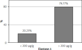
Pancreatic exocrine dysfunction was indicated in over 20% of patients because their faecal elastase-1 levels were below the reference value of < 200 µg/g. There was no significantly higher prevalence of reduced faecal elastase-1 levels in patients reporting abdominal pain (p = 0.997). However, patients with exocrine pancreatic insufficiency were significantly more likely to report the presence of diarrhoea (p = 0.002).
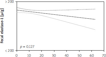
There was no significant effect of diabetes duration on elastase-1 levels in the patients examined. The pattern of interdependence between the two parameters was not statistically significant (p = 0.127).
The multivariate analysis of the parameters under analysis demonstrated that the only statistically significant coefficient (p £ 0.05) correlating with faecal FE-1 concentration (Table III) was elevated body mass index (BMI). No significant correlation was observed between reduced FE-1 concentration and the degree of diabetes compensation, age at diagnosis, type of diabetes, or type of anti-diabetic treatment used (Figures 1 and 2).


Discussion
Individuals within the diabetic patient population are particularly vulnerable to developing both mild and severe forms of PEI. Prior research, including smaller cohort studies, such as the preliminary Polish investigation, has suggested a significant correlation between both type 1 and type 2 diabetes mellitus (DM) and exocrine pancreatic dysfunction [7, 11, 13–19]. Noteworthy findings from studies, such as those conducted by Hardt et al., reveal that up to 40.7% of individuals with diabetes may experience exocrine pancreatic insufficiency [13]. In contrast, Vujasinovic et al. (2012) performed a single-centre analysis involving 150 patients with DM1 and DM2, identifying PEI in only 5.4% of cases, yielding results that deviate markedly from other similar studies. It is important to consider the possibility that the findings may be influenced by the implementation of exceedingly stringent inclusion criteria, potentially leading to an underrepresentation of reduced elastase levels within the Slovenian cohort [14]. Such disparities indicate that the prevalence of PEI in diabetic patients may significantly fluctuate depending on regional and demographic factors. The present study corroborates the observations made in the initial Polish study, demonstrating that reduced faecal elastase-1 (FE-1) levels (FE-1 < 200 µg/g) were present in 20.2% of all patients with diabetes combined (both type 1 and type 2). Therefore, routine evaluation of pancreatic exocrine function in diabetic patients is crucial in clinical settings.
FE-1 for detecting PEI has been shown to have a sensitivity of 54–90% and a specificity of 80% in mild to moderate PEI cases [20]. In severe PEI, the sensitivity of FEL-1 increases to 100% and specificity to 93% [20–25]. It is important to note that false-positive FE-1 results can occur in instances of stool contamination by urine or diarrhoea [26]. The utilisation of FE-1 for the diagnosis of PEI has been demonstrated to be less accurate in comparison to alternative testing methodologies, such as those involving faecal fat collection. However, FE-1 is widely available and is regarded as a satisfactory alternative. Furthermore, the execution of additional direct tests can present challenges for patients and staff members, and these tests can be both time-consuming and expensive [26, 27].
The initial Polish study explored the association between PEI and the occurrence of abdominal symptoms, but no statistically significant differences were noted between the prevalence of PEI and abdominal symptoms. However, a correlation between the presence of such symptoms and small intestinal bacterial overgrowth (SIBO) was confirmed [16]. This underscores the imperative for further investigations to comprehensively assess the prevalence and ramifications of PEI in diabetic patients.
The present study, conducted on a larger cohort of diabetic patients, documented a higher incidence of diarrhoea (24.9%) and abdominal pain (35.1%) compared to the previous Polish study [16], which recorded rates of diarrhoea at 17.9% and abdominal pain at 21.9%. This discrepancy might be indicative of a phenomenon specific to this population. Conversely, a literature review by Mohapatra et al. suggests that diabetic exocrine pancreatopathy is frequently asymptomatic [10]. When considering the hypothesis that PEI may be a late complication of diabetes, most existing literature fails to demonstrate a significant correlation between FE-1 levels and the duration of either DM1 or DM2 [7, 14, 28–30]. It is noteworthy that only one publication has indicated a correlation between diabetes duration and reduced FE-1 levels [13]. In alignment with this perspective, Hardt’s assertion regarding the association between reduced elastase-1 and diabetes duration lacks statistical significance, as acknowledged by the author, who mentioned that the correlation becomes significant only with a larger patient sample [13]. In our analysis, the linear regression analysis yielded no statistically significant results (p = 0.127), although a slight upward trend in PEI prevalence was observed with an increase in diabetes duration. These findings are consistent with prior literature, reinforcing the notion that pancreatic exocrine dysfunction is not merely a late complication of diabetes, but rather can be diagnosed irrespective of disease duration.
From a clinical standpoint, these findings are of considerable significance, underscoring the imperative for the assessment of PEI in individuals with DM1 and DM2, irrespective of disease duration or the presence of abdominal symptoms. The timely diagnosis of pancreatic exocrine insufficiency should be prioritised at the early stages of diabetes, thus advocating for the routine determination of FE-1. Early recognition of impaired pancreatic exocrine function is crucial for the timely initiation of enzyme replacement therapy, which is imperative for the management of PEI and the mitigation of associated complications, including malabsorption of fats, proteins, and vitamins, particularly vitamin D. Chronic vitamin D deficiency has been associated with an increased risk of osteoporosis and osteopaenia in diabetic patients [31–33].
Furthermore, the multivariate analysis revealed that the only statistically significant correlation (p < 0.05) with faecal FE-1 concentration was a higher BMI (Table III). No significant relationship was observed between reduced FE-1 levels and other factors, such as diabetes compensation level, age at diagnosis, diabetes type, or antidiabetic treatment methods.
It is noteworthy that 71.43% of the study participants exhibited a BMI of 25 or above, indicating that a considerable proportion of diabetic individuals in this cohort were classified as overweight. This outcome contrasts with those of earlier studies, including those by Cavalot et al. (2006), Hahn et al. (2008), Icks et al. (2011), and Larger et al. (2012), which did not identify any link between elevated BMI and PEI prevalence in diabetic patients [7, 17].
Among studies focusing on DM2 patients, only Yilmaztepe et al. (2005) failed to demonstrate an association between elevated BMI and pancreatic exocrine disorders. Conversely, Rathmann et al. (2001) and Larger et al. (2012) identified low BMI as a significant factor associated with PEI [7, 17, 28]. Furthermore, Rathmann et al. noted that low elastase-1 values were more prevalent among non-obese diabetic patients [28]. This study presents a divergent perspective on this matter. From a clinical standpoint, the observed lower BMI values may be attributed to malabsorption stemming from pre-existing pancreatic exocrine dysfunction. Thus, it is critical to evaluate patients with diabetes for pancreatic exocrine insufficiency independently of their BMI. It is noteworthy that individuals with a higher BMI are more prone to developing diabetes. In conclusion, a timely diagnosis of diabetic exocrine pancreatopathy is crucial to averting complications associated with PEI, which primarily manifest as malabsorption of fats, proteins, vitamins, and micronutrients [34–36]. Recognising this condition in a timely manner is imperative to enhance patient care and improve clinical outcomes.
Conclusions
This study highlights the high prevalence of exocrine pancreatic insufficiency in diabetic patients, emphasising the need for effective screening and management strategies. From a clinical perspective, it is recommended that diabetic patients be screened for elastase-1 concentration in their stool, and that those with a confirmed diagnosis of PEI be considered for enzyme replacement therapy. It is noteworthy that the incidence of diabetic pancreatopathy is independent of both the duration of diabetes and the type of treatment utilised.














