
Deficient fixation of midgut components can lead to varying degrees of midgut malrotation [1]. The malrotated bowel is at risk of torsion, resulting in midgut volvulus. In many cases, intestinal malrotation remains undetected or asymptomatic. Traditionally, the diagnosis of midgut volvulus relied on upper gastrointestinal (GI) fluoroscopy series, while ultrasonography was not recommended as a first-line imaging modality. However, grey-scale and Doppler ultrasound can reveal a reversed relationship between the superior mesenteric artery (SMA) and the superior mesenteric vein (SMV), which is commonly associated with intestinal malrotation [2]. Furthermore, the “whirlpool sign” on abdominal ultrasound documents – accurately and without exposure to radiation – a clockwise wrapping of the mesentery and the superior mesenteric vein around the superior mesenteric artery, which has a strong predictive power for volvulus [3]. This report presents a case of neonatal midgut malrotation complicated by volvulus, diagnosed solely via ultrasound, allowing prompt surgical intervention to eliminate the risk of bowel ischaemia and subsequent necrosis. Initial sonographic findings that facilitated the diagnosis and guided the treatment of this complex condition, without the need for upper GI fluoroscopy, are detailed. A 29-day-old, full-term neonate, was admitted to the emergency ward of a tertiary care centre with a history of non-projectile vomiting since day 17 after birth. Based on medical records, the vomiting episodes initially occurred every 1 to 2 days but had gradually increased daily over the past 3 days. On the day of admission, the infant experienced three episodes of bilious vomiting. Physical examination revealed abdominal distention, agitation, poor feeding, mild tachycardia (172 bpm), and reduced urination. Upon admission (after 6 h of the patient’s transportation), a nasogastric tube was placed to relieve the stomach. Additionally, issues such as dehydration and electrolyte imbalance were immediately corrected. An abdominal X-ray revealed mild stomach distention with air in the first portion of the duodenum (Figure 1). Without any further delay, abdominal ultrasound was performed within the first hour of the baby’s admission, by a paediatric sonographer using a high-resolution, linear 9–15-MHz probe (Logiq E10 Series; GE, Milwauke, WI, USA). No signs of hypertrophic pyloric stenosis were present (normal transverse muscle diameter, measuring approximately 0.24 cm). Investigation of the relationship of the SMV and the SMA, and the third section of the duodenum was required following examination of the pylorus. Colour Doppler ultrasound documented an inverted relationship between SMA and SMV, compatible with intestinal malrotation. A routine full abdomen scan in the supine position followed by a 4-quadrant bowel survey revealed a spinning cluster of small bowel loops around the superior mesenteric artery depicted on transverse views, forming a “whirlpool” configuration, both on A-B mode and colour Doppler images (PRF: 2, 3; Wall Filter: 251). This finding was highly indicative of midgut volvulus (Figures 2 A, B). In addition, a portion of the 3rd part of the duodenum was only partially visualised (owing to the baby’s continuous crying) just in front of the superior mesenteric artery, as a clue to its intraperitoneal location (Figure 3). Due to the baby’s deteriorating condition and diagnostic assurance from the US results, informed consent was obtained from the parents before proceeding with surgical exploration. A typical Ladd’s procedure was performed. When entering the abdomen, the caecum was at the right upper quadrant, and loops of the terminal ileum were found twisted 180° in an anticlockwise manner (Figure 4). An intraperitoneal position of the 3rd part of the duodenum was confirmed. The blood supply, although compromised due to the bowel torsion, did not seem to have caused any serious damage to the bowel loops, which were viable and functioning. At that point, the twisted bowel loops were brought to a normal position by manually rotating the terminal ileus clockwise, and a typical Ladd’s procedure was performed, which included dividing all the obstructing bands, broadening the mesentery base, and relocating the small intestine to relieve tension and restore intestinal patency. The mesentery base initially was measured at 26 mm width, and after the Ladd’s procedure it measured 39 mm. Finally, a prophylactic appendicectomy was performed. The newborn was fed 24 h after the operation, initially with 30 ml formula milk, which was increased by 10 ml every 2 h up to 80 ml, and after that she was freely breast fed. She had an uneventful postoperative course and was discharged on the fifth postoperative day. Follow-up at 1 month showed no complications.
Figure 1
Inverted relationship between the superior mesenteric artery (SMA – thin arrow) and superior mesenteric vein (SMV – thick arrow) on grey-scale ultrasound
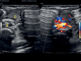
Figure 2
B-mode (A) and colour Doppler (B) ultrasound images illustrating the “whirlpool sign” (yellow arrow) – a clockwise rotation of the small bowel and superior mesenteric vein around the centrally located superior mesenteric artery
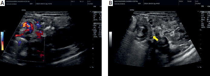
Figure 3
The third portion of the duodenum was not visualised behind the SMA but was only partially depicted as a grey longitudinal area just in front of the orifice of the SMA (yellow empty arrow)
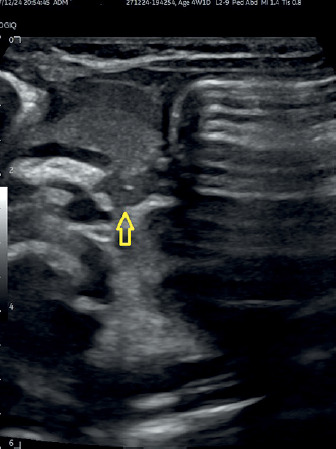
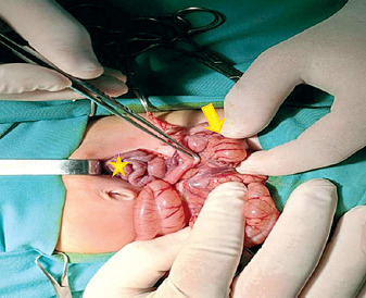
Figure 4
Surgical intervention findings – arrow indicates the cecum, and asterisk marks the twisted small intestinal loops
The outcome of babies who undergo Ladd’s procedure due to malrotation and volvulus generally depends on the duration of symptoms and the underlying pathology caused by the twisted bowel and the compromised blood supply. Initiation of feeds is strongly dependent on the condition of the bowel after the operation and whether there was a bowel resection performed. Luckily, in this case the twisted bowel was viable and functioning, which allowed early initiation of feeding. However, it is obvious that the timing of discharge also depends on the surgical findings. Nevertheless, even when a short segment of bowel is resected, the initiation of feeding and thus the time of discharge is rarely more than 7 postoperative days. Things become more complicated if a large segment of bowel has to be resected.
Congenital intestinal malrotation occurs in 1 in 200 to 500 live births. While many individuals remain asymptomatic throughout their lives, this condition can lead to serious complications such as midgut volvulus [1]. Anomalous intestinal rotations can present in various forms, all stemming from an interrupted rotation of the duodenum and/or caecum around the SMA axis during embryologic development. The intestine, when abnormally rotated and fixated, is suspended from a narrow mesenteric base, increasing its susceptibility to torsion and volvulus [4]. If blood flow is compromised, it can result in life-threatening bowel infarction.
Systematic physical examination, and antenatal and postnatal clinical records may assist in the differential diagnosis of bilious vomiting by indicating cases of medical origin with no need for further exploration. In the absence of evidence to the contrary, bilious vomiting is considered to be the result of an underlying surgical pathology, invariably due to intestinal obstruction. Amongst these surgical causes, only 3–5% of cases of bilious vomiting present on the grounds of malrotation with volvulus. Despite its low incidence, it remains a time-critical and life-threatening condition [5].
In symptomatic malrotation (with or without volvulus), affected neonates usually present in the first week of life in approximately 75% of cases and in 15% during the first year of life. The remaining 10% of cases may present at any age [5]. Bilious emesis occurs either due to obstruction secondary to volvulus or partial obstruction due to Ladd’s bands causing extrinsic compression of the duodenum [5]. Conversely, in our case, symptoms were nonspecific initially, with visible bilious content not during the first week of life but only on the day of admission. In cases of partial obstruction, vomiting may occur occasionally, containing bile, but without abdominal distention. If torsion is loose, the patient may experience intermittent pain. However, in cases of complete duodenal blockage and torsion, as in our case, biliary vomiting continues, accompanied by abdominal distention, dehydration, weight loss, and irritability. This situation increases the risk of small intestinal necrosis, peritonitis, or even sepsis [6]. Midgut volvulus is a significant paediatric emergency that often presents serious diagnostic challenges. Abdominal X-ray may indicate duodenal obstruction by showing a dilated stomach and duodenum, but up to 20% of cases appear normal [7–9]. Even with air present in the duodenum and colon, malrotation and volvulus cannot be definitively ruled out, complicating the differential diagnosis. Until recently, diagnosing intestinal malrotation and volvulus primarily relied on upper gastrointestinal contrast-enhanced series. These studies help visualise the abnormal fixation of the duodenojejunal junction and caecum [9]. Barium studies can show signs of malrotation-volvulus, such as a dilated duodenum and proximal small bowel obstruction, or the characteristic “corkscrew” configuration. However, the presence of the corkscrew sign is not always definitive, especially in cases of atypical or partial bowel rotation [6]. Meanwhile, X-ray studies have certain limitations for neonates, such as radiation risks, problems in the patient’s positioning, and not being time effective [10]. Moreover, routine UGI series may not be needed for neonates with non-surgical bilious emesis, and they present with an up to 30% false positive rate in the imaging workup for malrotation [10]. Interestingly, the use of abdominal ultrasound in neonates with bilious vomiting has been rapidly increasing in recent years as an alternative to upper gastrointestinal contrast series [5, 10]. Ultrasonography (US) combined with colour Doppler can reveal a reversed relationship between the SMA and SMV, which is suggestive of malrotation [11]. A key advantage of ultrasound is that it has no radiation exposure, which is crucial for children and for maintaining radiation protection principles [6]. In a systematic review and meta-analysis by Nguyen et al. [12] including 17 cohort or cross-sectional studies and 2257 participants, a summary sensitivity of 94% (95% CI: 89–97%) and summary specificity of 100% (95% CI: 97–100%) (moderate certainty evidence) were derived for the use of ultrasound in the diagnosis of malrotation with or without midgut volvulus.
In cases where malrotation is complicated by small intestine volvulus, the “whirlpool sign” can be observed on colour Doppler ultrasound. This sign indicates a clockwise wrapping configuration of small intestine loops, the mesentery, and the SMV surrounding the SMA, directly correlating the anatomical abnormalities seen in midgut volvulus [5, 13]. In the case presented the infant’s history of deteriorating emesis, critical condition upon admission, and the presence of the “whirlpool sign” on ultrasound made it unnecessary to perform an upper GI tract series, leading to timely surgical intervention. Nguyen et al., in their most recent multicentre, retrospective, cross-sectional study [14], showed that abdominal ultrasound for midgut volvulus presented with sensitivity and specificity of 97% and 98%, respectively, by original report and 97% and 99%, respectively, by blinded research review. Specific sonographic signs such as the whirlpool sign exhibited 99% accuracy for diagnosing of volvulus. In the same study, Nguyen et al. also highlighted the presence of the intraperitoneal third part of the duodenum as a more specific sign for intestinal malrotation, with a reported accuracy of 98% [14]. In malrotation, the duodenum may be abnormally fixated, such as being intraperitoneal instead of retroperitoneal, which can lead to complications such as volvulus due to this abnormal position of the bowel. Detecting these positional changes by ultrasound is crucial for diagnosing malrotation and preventing significant gastrointestinal complications.
Several study groups of an expert panel narrative review in 2022 conducted by Nguyen et al. support the administration of normal saline or sterile water via an enteric tube as an alternative to better delineate the duodenal course, using the fluid-filled stomach as a sonographic window and following the bolus of fluid through the duodenum [15].
There is some scepticism about the role of abdominal ultrasound US due to the detrimental consequences of a potentially missed diagnosis of volvulus. Reported false-positive and false-negative rates of abdominal ultrasound are 0% and 6%, respectively. However, false-negative results for both malrotation and volvulus may also occur for upper gastrointestinal contrast-enhanced series [15]. Alehossein et al. [10] also suggested that ultrasound can effectively investigate the surgical cause of bilious emesis in neonates, thus obviating the drawbacks of irradiation, cost, and time, especially in life-threatening conditions such as midgut volvulus.
Nonetheless, abdominal ultrasound can successfully exclude other differentials of bilious emesis, such as hypertrophic pyloric stenosis, as in our case, whereas the pyloric muscle dimensions were within normal limits. Additionally, other pathologies related to neonatal vomiting including intussusception can be revealed via ultrasound by the depiction of the classic “donut” or “target” signs, which refer to a central echogenic ring of fat within a round soft tissue mass [16].
In conclusion, ultrasound is an essential tool for diagnosing midgut malrotation and volvulus. It can identify specific imaging signs, such as the reversed orientation of the SMA and SMV, and the intraperitoneal position of the third part of the duodenum. Most importantly, it can reveal the “whirlpool sign”, as shown in our case. US as a cost-effective imaging modality enables prompt surgery in neonates, minimising delays and radiation exposure. Upper gastrointestinal (UGI) series should only be used in cases where ultrasound results are nondiagnostic or equivocal.














