eISSN: 1897-4317
ISSN: 1895-5770
Gastroenterology Review/Przegląd Gastroenterologiczny
Bieżący numer Archiwum Artykuły zaakceptowane O czasopiśmie Rada naukowa Bazy indeksacyjne Prenumerata Kontakt Zasady publikacji prac
Panel Redakcyjny
Zgłaszanie i recenzowanie prac online
NOWOŚĆ
Portal dla gastroenterologów!
www.egastroenterologia.pl
SCImago Journal & Country Rank
1/2022
vol. 17
 
Poleć ten artykuł:
Udostępnij:
List do Redakcji

A rare variant of ileus – Bouveret’s syndrome

Weronika Frąk
1
,
Adam Durczyński
2
,
Piotr Hogendorf
2
,
Adam Fabisiak
1
,
Ewa Małecka-Wojciesko
1

1.
Department of Digestive Tract Diseases, Faculty of Medicine, Medical University of Lodz, Lodz, Poland
2.
Department of General and Transplant Surgery, Faculty of Medicine, Medical University of Lodz, Lodz, Poland
Gastroenterology Rev 2022; 17 (1): 83–84
Data publikacji online: 2022/03/18
Plik artykułu:
- A rare variant.pdf  [0.09 MB]
Pobierz cytowanie
 
Metryki PlumX:
 

Mechanical small bowel ileus may have several causes. In this Letter to the Editor we would like to remind of its rare cause, i.e. the gallstone ileus – Bouveret’s syndrome, based on our case. Bouveret’s syndrome is a relatively rare variant of intestinal obstruction [1] caused by a gallstone migrating through the bilio-duodenal fistula. Proper treatment can be delayed due to the nonspecific symptoms, which may cause a considerable mortality [2, 3]. Radiological signs, known as Rigler’s triad for gallstone ileus, include pneumobilia, an ectopic gallstone, and small bowel obstruction. Imaging of the abdomen by plain X-rays or computer tomography (CT) is recommended [4]. The mainstay of management is surgical removal of the impacted stone [5].

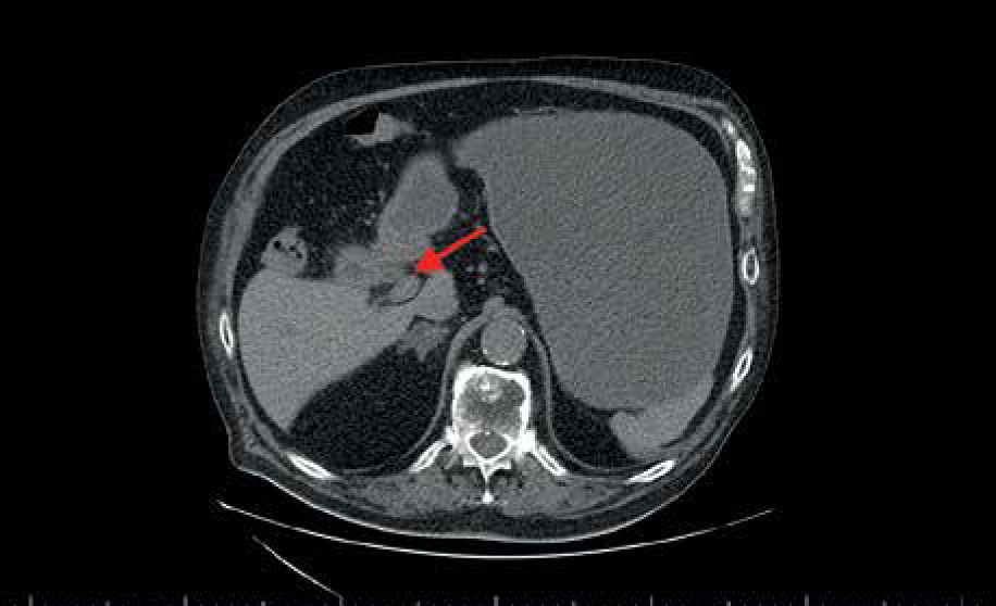
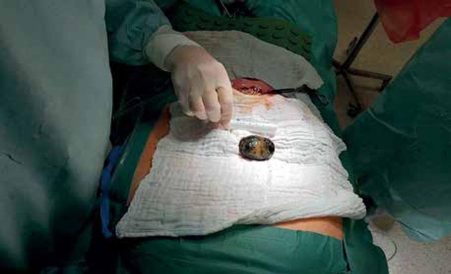
A 79-year-old man presented to the Emergency Department with a 5-day history of abdominal pain and persistent vomiting. His medical history included a type 2 diabetes mellitus, hypertension, cholecystolithiasis, and obesity. His initial laboratory evaluation revealed a high white blood cell count (15.3 × 10³/µl) and elevated C-reactive protein (44.3 mg/l). The patient was referred for an abdominal CT scan, and radiological findings suggested a small bowel obstruction. CT showed a distended, liquid-filled stomach and the ectopic gallstone in the proximal jejunum, which appeared large and round (Figure 1). Proximal small bowel loops were liquid filled and dilatated and distally collapsed. In addition, the thickening of the gallbladder wall, pericholecystic fat stranding, pneumobilia, and bilio-duodenal fistula were visualized (Figure 2). The patient underwent an emergency surgery, and a 6-cm stone was successfully retrieved (Figure 3). The surgery and initial postoperative course have been uneventful.
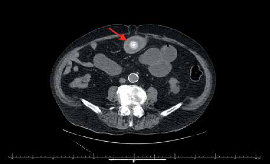
Figure 1

Abdominal CT. The red arrow points to the ectopic gallstone in the first part of the duodenum

/f/fulltexts/PG/46633/PG-17-46633-g001_min.jpg
Figure 2

Abdominal CT. The red arrow points to the bilio-duodenal fistula

/f/fulltexts/PG/46633/PG-17-46633-g002_min.jpg
Figure 3

A 6-cm-long retrieved gallstone

/f/fulltexts/PG/46633/PG-17-46633-g003_min.jpg

Bouveret’s syndrome is rare and represents less than 5% of all cases of gallstone ileus [6]. The common risk factors are female sex, older age, with frequent associated comorbidities, obesity, and gallstones size greater than 2.5 cm. Common clinical symptoms are abdominal pain, nausea, and vomiting. Rigler’s triad in radiologic examination are necessary to confirm the diagnosis. Although, there are no definitive guidelines on the approach and management of this condition, the most common is surgical treatment [7]. The tailored procedure must be considered individually, based on the patient’s condition and age. Moreover, optimal time of surgery and sometimes a restrictive surgical approach are crucial for satisfactory patient outcomes.

Acknowledgments

This work was supported by a grant from the Medical University of Lodz (503/1-002-01/503-11-001-19-00 to EMP)

Conflict of interest

The authors declare no conflict of interest.

References

1 

Haddad FG, Mansour W, Deeb L. Bouveret’s syndrome: literature review. Cureus 2018; 10: e2299.

2 

Chow BL, Zia K, Scott S, et al. The curious case of biliary emesis and bowel obstruction from Bouveret syndrome BMJ Case Rep 2019; 12: e230194.

3 

Enumah ZO, Wong EG, Kent AJ. Complicating factors in the management of advanced Bouveret syndrome in frail and medically complex patients: case report and discussion of pathophysiology. Int J Surg Case Rep 2020; 77: 96-9.

4 

Nickel F, Müller-Eschner MM, Chu J, et al. Bouveret’s syndrome: presentation of two cases with review of the literature and development of a surgical treatment strategy. BMC Surg 2013; 13: 33.

5 

Singh G, Merali N, Shirol S, et al. A case report and review of the literature of Bouveret syndrome. Ann R Coll Surg Engl 2020; 102: e15-9.

6 

Osman K, Maselli D, Kendi AT, et al. Bouveret’s syndrome and cholecystogastric fistula: a case-report and review of the literature. Clin J Gastroenterol 2020; 13: 527-31.

7 

Al-Habbal Y, Ng M, Bird D, et al. Uncommon presentation of a common disease–Bouveret’s syndrome: a case report and systematic literature review. World J Gastrointest Surg 2017; 9: 25-36.

Copyright: © 2022 Termedia Sp. z o. o. This is an Open Access article distributed under the terms of the Creative Commons Attribution-NonCommercial-ShareAlike 4.0 International (CC BY-NC-SA 4.0) License (http://creativecommons.org/licenses/by-nc-sa/4.0/), allowing third parties to copy and redistribute the material in any medium or format and to remix, transform, and build upon the material, provided the original work is properly cited and states its license.
 
© 2024 Termedia Sp. z o.o.
Developed by Bentus.