Introduction
Nonsteroidal anti-inflammatory drug (NSAID)-exacerbated respiratory disease (N-ERD) is a chronic inflammatory disorder of the respiratory tract. It occurs in patients with asthma and/or chronic rhinosinusitis with nasal polyps (CRSwNP), whose symptoms are exacerbated by strong cyclooxygenase 1 (COX-1) inhibitors, including aspirin and other NSAIDs [1]. The disease affects approximately 7–10% of adult asthmatics, but its prevalence rises to 15% in patients with severe asthma [2]. This condition has been reported in 8-26% of patients with CRSwNP [3]. The presence of CRSwNP in patients with N-ERD ranges from 60–99% [4, 5]. Patients with N-ERD tend to have moderate to severe asthma with frequent exacerbations, and more often require oral corticosteroids and higher doses of inhaled corticosteroids to control the disease compared to NSAIDs-tolerant asthmatics [6]. These patients also have more severe sino-nasal disease requiring repeated sinus surgeries and oral corticosteroid bursts compared to patients with CRSwNP alone [7].
The fundamental pathomechanism of N-ERD is dysregulation of arachidonic acid metabolism, which in turn leads to constant overproduction of the proinflammatory and bronchoconstrictor cysteinyl leukotrienes (LTs) – mainly leukotriene E4 (LTE4) and prostaglandin D2 (PGD2) (Figure 1). Along with this comes underproduction of anti-inflammatory cyclooxygenase-2 (COX-2), prostaglandin E2 (PGE2), and membrane-bound PGE2 synthase-1 (mPGE-1), and diminished expression of the receptor for PGE2 (EP2) [8]. Thus, the core problem in N-ERD is not solely an excess of proinflammatory mediators but also a deficiency of anti-inflammatory regulators, including PGE2, which normally plays a crucial role in inhibiting mast cell and eosinophil activation, as well as LTs secretion [8]. Aspirin and other NSAIDs exert their effects by irreversible inhibition of COX-1, while in N-ERD this inhibition further reduces the already deficient PGE2 production and shunts arachidonic acid towards the 5-lipooxygenase (5-LOX) pathway. This together leads to an acute, exaggerated rise in proinflammatory LTs and PGD2 [9]. This acute biochemical imbalance precipitates the characteristic hypersensitivity reactions to aspirin and other COX-1 inhibitors.
FIGURE 1
Overview of the pathogenetic mechanisms of N-ERD and potential targets of available biologics ASA – acetylsalicylic acid, COX-2 – cyclooxygenase 2, EP2 – prostaglandin E2 receptor, ILC2 – innate lymphoid cells, IL-4 – interleukin 4, IL-5 – interleukin 5, IL-13 – interleukin 13, LTs – leukotrienes, mPGES-1 – membrane-bound PGE2 synthase-1, PGD2 – prostaglandin D2, PGE2 – prostaglandin E2, Th2 – lymphocytes Th2.
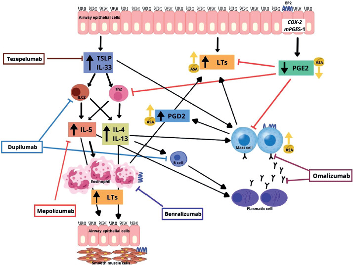
From a histological point of view N-ERD is characterised by an intense, ongoing eosinophilic inflammation in the upper and lower airways and frequent presence of degranulated mast cells [10]. Most studies have shown a cytokine and chemokine milieu consistent with type 2 inflammation with the involvement of such cytokines as IL-4, IL-5, IL-13, and alarmins (IL-33, IL-25, TSLP) [8]. However, a recent study carried out at the level of the upper and lower airways suggests a more heterogeneous inflammatory profile of N-ERD with involvement of inflammation other than type 2 [11].
Because many patients with N-ERD have severe asthma and/or severe CRSwNP, there is an evident indication for the use of biological therapy in this cohort. Post hoc analyses of the pivotal studies have shown that this group of patients benefited equally from the biologic treatment as those patients who were NSAIDs-tolerant [12–14]. However, it should be noted that in these studies the diagnosis of N-ERD was made on the basis of medical history alone. There are few studies carried out in well-defined, confirmed by aspirin challenge groups of patients with N-ERD that have attempted to investigate the effect of biologics on the pathomechanisms of the disease, and especially their role in restoring aspirin tolerance.
The effects of biologics in patients with N-ERD
Omalizumab
Hayashi et al. [15] prospectively evaluated 21 allergic patients with severe N-ERD confirmed by oral aspirin challenge, treated with omalizumab for 12 months. According to the Global Evaluation of Treatment Effectiveness (GETE) scale, 18 of 21 patients responded positively for the treatment. The number of exacerbations and hospitalisations, as well as daily doses of systemic corticosteroids, were significantly reduced. Respiratory function, nasal- and asthma-related symptom scores improved significantly. Also, omalizumab significantly reduced blood eosinophil counts (BEC), and urinary concentrations of leukotriene E4 (LTE4) and prostaglandin D2 metabolite (PGD2M) as assessed before and after omalizumab treatment. It was the first report to demonstrate that omalizumab displayed rapid clinical effectiveness and inhibited mast cell activation, as well as LTE4 and PGD2M overproduction in N-ERD.
The same research group later performed the first randomised, double-blind, placebo-controlled crossover study in a group of 16 allergic N-ERD patients receiving omalizumab or placebo in two treatment phases of 3-month duration with an 18-week washout period [16]. N-ERD was diagnosed by a positive result for oral aspirin challenge within the last 2 years before enrolment. Omalizumab treatment, in contrast to placebo, resulted in a rapid improvement of asthma and sino-nasal symptoms. Statistically significant improvements in scales evaluating asthma control and Sino-Nasal Outcome Test (SNOT-22) were observed at the first month of treatment and maintained in the following month. In the omalizumab treatment phase, compared to placebo, significantly lower urinary concentrations of LTE4 and tetranor-PGDM were observed since the first day. In the case of LTE4, significant differences persisted for 3 months, while concentrations of tetranor-PGDM also tended to be lower, although they did not reach statistical significance at the second and third month. The unique value of the study was an oral aspirin challenge, which was performed at the end of each treatment phase. At the end of the placebo phase, all patients showed a positive reaction to aspirin. Conversely, at the end of omalizumab phase, 10 of 16 (62.5%) patients had a negative reaction after reaching the highest cumulative aspirin dose, which indicated that these patients achieved complete aspirin tolerance. In 4 (25%) patients with a positive reaction, the cumulative aspirin dose increased when compared with the dose in the placebo phase, indicating partial improvement of tolerance, whereas in the other 2 patients the cumulative aspirin dose remained the same. Moreover, asthma symptoms and nasal symptoms were significantly milder in the omalizumab phase than in the placebo phase. Out of blood biomarkers, BEC, eosinophilic cationic protein (ECP), periostin, and tryptase were significantly lower in the omalizumab phase than in the placebo phase. The important point of the study was the measurement of urinary LTE4 and tetranor-PGDM concentrations before and during 24 hours of oral aspirin challenge. They were expressed as the area under the logarithm level of these biomarkers concentrations versus time curve (AUC[before-24h]) between the placebo and omalizumab phase, which reflected the overall eicosanoid production in response to systemic aspirin exposure. There was a significant reduction in the urinary concentrations of both eicosanoids in the omalizumab phase compared with the placebo phase. Moreover, the concentrations of LTE4 and tetranor-PGDM at each time point within 24 h after the aspirin challenge were significantly lower in the omalizumab phase than in the placebo phase. The results of this study suggest that omalizumab has inhibitory effect on mast cell function during an aspirin challenge and potentially during clinical adverse responses to aspirin. The fact that omalizumab significantly suppressed the urinary concentrations of LTE4 and tetranor-PGDM during treatment periods, when there was no aspirin exposure, suggests that it also has inhibitory effect on ongoing mast cell activation, one of the key players in the pathogenesis of N-ERD.
In the prospective trial, Quint et al. [17] went further and investigated the effect of omalizumab treatment on aspirin tolerance in atopic and non-atopic patients with N-ERD as examined by oral aspirin challenge at baseline and after 6 months of treatment. Out of 33 patients included in the study, 15 (56%) developed complete aspirin tolerance, 5 (19%) tolerated higher aspirin doses, and 7 (26%) did not show any symptom improvement, reacting to aspirin doses of 125 mg or 250 mg. None of the patients experienced a worsening of symptoms after re-exposure to aspirin. Overall clinical effectiveness of omalizumab was very good. Polyp size and symptoms of CRSwNP were significantly reduced, as evaluated by nasal polyp score and SNOT 22. Asthma control test (ACT) revealed a significant improvement of subjective pulmonary symptoms, while spirometric data demonstrated a mild improvement. The comparison between the atopic and non-atopic group showed that omalizumab-induced complete aspirin tolerance and clinical efficacy in the treatment of CRSwNP and asthma was independent of atopic sensitisation. It should be assumed that concomitant allergic sensitisation is irrelevant for successful omalizumab therapy and restoring aspirin tolerance in N-ERD patients. On the other hand, patients with omalizumab-induced aspirin tolerance showed a trend in higher levels of serum IgE, ECP, and an elevated relative number of eosinophils at baseline compared with intolerant patients. It suggests that these biomarkers might be indicative of omalizumab response in this population.
In another study, Hayashi et al. [18] investigated the clinical efficacy of omalizumab against N-ERD-related extra-respiratory symptoms at a steady state, without aspirin exposure and during oral aspirin challenge. In the retrospective, observational part of the study 27 patients were evaluated with respect to extra-respiratory episodic N-ERD-related symptoms such as chest pain, and gastrointestinal and cutaneous symptoms. During a 1-year omalizumab treatment, the frequency of patients experiencing extra-respiratory symptoms and the number of exacerbations of these symptoms were significantly reduced. Omalizumab treatment was also associated with a significant reduction in urinary LTE4 concentration. In the second part of the study, 3 N-ERD patients with aspirin challenge-induced extra-respiratory symptoms who participated in the previous randomised, placebo-controlled study [16] were investigated. In the omalizumab phase the following was observed: 1 patient did not develop any symptoms, in the second patient the dose of aspirin required to induce extra-respiratory symptoms increased, and in the third patient no extra-respiratory symptoms were induced at the same aspirin dose that provoked these symptoms in the placebo phase. Thus, the study demonstrated that omalizumab significantly ameliorated extra-respiratory symptoms not only at baseline conditions, but also during aspirin challenge. Concomitant reduction of urinary LTE4 concentrations suggests that omalizumab stabilises N-ERD by suppressing mast cell activation.
The results of the above-mentioned studies suggest that omalizumab may interfere with the pathomechanisms of N-ERD by its influence on mast cell function (Figure 1). They also indicate the potential role of IgE in the disease. Dysregulation of mast cell activation and overproduction of LTE4 and PGD2 by these cells is one of the pivotal components of N-ERD [19]. Because omalizumab suppresses free IgE and reduces the expression of high-affinity IgE Fc receptors on mast cells, it inhibits mast cell activity and consequently eosinophilic inflammation. This may explain the decrease in urinary LTE4 and PGD2 metabolite concentrations during omalizumab treatment observed in N-ERD patients in resting conditions [15, 16, 18] when ongoing mast cell activation is present [10]. The inhibitory effect of omalizumab on eicosanoids production during aspirin challenge, and potentially during clinical adverse response to aspirin, is particularly important [16]. These results, along with the observation that omalizumab treatment can restore aspirin tolerance in some N-ERD patients are particularly promising [16]. If confirmed in larger groups of patients, omalizumab could become a drug modifying N-ERD.
Dupilumab
Buchheit et al. [20] prospectively observed 22 patients with physician-diagnosed N-ERD who were treated with dupilumab as an add-on to severe asthma or CRSwNP therapy for 3 months. Eight participants were taking daily aspirin therapy after desensitisation for at least 6 months before starting dupilumab treatment, but they did not achieve sufficient control of asthma or CRSwNP. After initiating dupilumab, these patients had rapid improvement in clinical measures, including sense of smell, sino-nasal symptoms, asthma control, and lung function. The improvement of symptoms was observed just after the first month of treatment, and they were even better when measured after 3 months. Eicosanoids concentrations were measured in urine and nasal fluid. Urinary LTE4 concentrations decreased significantly after 1 and 3 months of dupilumab treatment, but there were no significant changes in urinary concentrations of PGD2 metabolite (tetranor PGD-M), although there was a trend towards a decrease at month 1. Urinary concentrations of prostaglandin E2 metabolite (PGE-M) were unchanged. Similarly to urine, nasal LTE4 concentrations significantly decreased at both time points when compared to baseline. In contrast, there were no dupilumab-induced changes in nasal concentrations of PGD2 metabolite (DHKPGD2), but a significant increase in concentrations of nasal fluid PGE2 was observed. Baseline severity of smell loss was correlated with lower PGE2 concentrations.
In a prospective open-label study, Schneider et al. [21] observed 31 N-ERD patients with hypersensitivity to this drug previously confirmed by oral aspirin challenge and then treated with dupilumab. The therapy, lasting 6 months, resulted in a significant reduction of polyp size, a decrease in the total SNOT-22 score, and improvement in smell perception and asthma symptoms measured by ACT. The lung function parameters showed a tendency to improve. Urinary concentrations of LTE4 and 11β-prostaglandin F2α showed a significant reduction. Dupilumab treatment was associated with an increase in absolute and percentage eosinophil blood levels and eosinophil cationic protein, and a decrease in total serum IgE levels. In addition, a drop in total IgE levels in nasal secretions was observed. Among other biomarkers assessed in nasal secretions, a significant decrease in type 2-associated cytokines and inflammatory cytokines was observed. Out of 31 patients included in the study, 30 completed the second aspirin challenge after 6 months of dupilumab treatment. Seven (23%) patients developed complete aspirin tolerance, and 10 (33%) patients reacted to higher doses than before treatment, demonstrating partial improvement in aspirin tolerance. Moreover, at the second aspirin challenge, patients mainly experienced isolated upper respiratory tract reactions, and fewer patients experienced both upper and lower respiratory tract reactions when compared with baseline challenge.
Taken as a whole, the aforementioned results suggest that dupilumab treatment may restore the disturbed balance of eicosanoid metabolism in N-ERD and influence the pathogenic mechanisms of the disease. The beneficial effect of dupilumab on the eicosanoid profile and, consequently, on clinical symptoms in patients with N-ERD also suggests that this drug interferes with the pathomechanisms of the disease (Figure 1). IL-4 is one of the cytokines present in the inflammatory milieu in N-ERD and plays a role in the dysregulation of COX pathway [22]. It can contribute to an increase in LTE4 synthase (LTE4S) expression and can inhibit the expression of mPGE-1, COX-2, and consequently PGE2 release, which is a typical phenomenon in N-ERD [23]. It has been hypothesised that dupilumab, by blocking the action of IL-4, restores normal function of the so-called the autocrine metabolic loop, consisting of COX-2, PGE2, EP2, and mPGE-1, and in this way, it normalises EP2 and COX-2 expression and the synthesis of both PGE2 and LTs [24]. This hypothesis may explain the decrease in urinary and nasal LTE4 concentrations along with the increase in nasal PGE2 concentration in N-ERD patients treated with dupilumab [20]. The lack of a simultaneous dupilumab-induced decrease in urinary and nasal PGD2 concentrations remains an open question [20]. Possibly cells other than mast cells are the main targets for dupilumab, considering the widespread IL-4Rα expression on multiple cell types. The restoration of autocrine metabolic loop by dupilumab may also explain the improvement of aspirin tolerance in N-ERD patients [21]. If these positive results are confirmed in larger groups of patients, dupilumab may also prove to be a disease-modifying drug. At the moment, it remains a mystery why the beneficial effect of biologics is observed in a subset of patients. It is possible that IL-4-mediated augmentation of LTs synthesis is more prevalent in some patients while in others LTs synthesis is more dependent on IgE-mediated pathway activation [22–24].
Mepolizumab
Studies on the effect of mepolizumab treatment on NSAID hypersensitivity in N-ERD patients are scarce and do not show such beneficial effects as therapy with omalizumab and dupilumab. Only a case series published by Martin et al. [25] demonstrated that mepolizumab does not prevent aspirin-induced reactions in patients with N-ERD. All three studied patients developed clinical reactions during oral aspirin challenge/desensitisation while on mepolizumab. The first patient, after 2 endoscopic sinus surgeries (ESS), since starting mepolizumab treatment reported mild improvement in asthma but continued to have nasal symptoms. After 3 months of mepolizumab treatment, she started desensitisation with aspirin, during which nasal blockage, rhinorrhoea, headache, and a drop of FEV1 by 12% from baseline were observed. The symptoms were induced with an aspirin dose of 162 mg. The second patient, after 4 ESS, since starting mepolizumab treatment reported fewer asthma exacerbations but continued to have poor asthma control and nasal symptoms. After 13 months of mepolizumab treatment, she started aspirin desensitisation, during which she developed pruritus, hives, and audible wheezing without a decrease in FEV1. The provocative dose of aspirin inducing these symptoms was 40 mg. The third patient, with 1 previous ESS, reported very little symptomatic improvement from the mepolizumab treatment and had continued anosmia. She started aspirin desensitisation after 6 months of such treatment, but she developed pruritus, severe abdominal pain, nausea, vomiting, and wheezing, with a decrease of FEV1 by 11% from baseline. The provocative dose of aspirin was 40.5 mg. These 3 cases showed that, regardless of the duration of mepolizumab treatment, patients with N-ERD can still develop aspirin-induced reactions. Therefore, continued aspirin and other NSAIDs avoidance is recommended unless the patients have successfully completed an aspirin desensitisation.
It can be assumed that although the tissue eosinophilia in most patients with N-ERD and CRSwNP is substantial, eosinophils are not the main effector cells that drive ongoing inflammation. Other effector cells, including mast cells, basophils, and epithelial cells, may play a key role in the disease because they also express IL-5Rα. Taken together, the response to biologic therapy in N-ERD patients is as complex as the pathogenesis of the disease itself.
Comparison of all biologics
So far, only one head-to-head study comparing the effect of biologic therapies on the induction of NSAID tolerance in N-ERD patients has been performed. Sanchez et al. [26] carried out a prospective pilot study in a real-world clinical setting among subjects with severe asthma, NSAID intolerance confirmed by oral aspirin challenge, and type 2 inflammation. A total of 38 patients were randomly allocated to 6-month treatment with one of the following biologics: benralizumab, dupilumab, mepolizumab, or omalizumab. There were no differences in terms of paraclinical and clinical characteristics, so they were able to receive any of the four biologic therapies. After completion of the study, the second oral aspirin challenge was performed. Compared to the baseline, only patients treated with omalizumab and dupilumab tolerated significantly higher doses of aspirin, while in patients treated with mepolizumab and benralizumab, changes in aspirin dose were not significantly different. The number of patients treated with omalizumab (n = 6/10, 60%) and dupilumab (n = 4/10, 40%) who achieved complete aspirin tolerance was higher than those treated with mepolizumab (n = 2/9, 22%) and benralizumab (2/9, 22%). The difference was statistically significant in favour of omalizumab and dupilumab over mepolizumab and benralizumab. No prognostic factors were found in terms of laboratory (BEC, total IgE) and clinical parameters that could be responsible for better aspirin tolerance in these patients. The results of this study are consistent with previously presented studies with a single biologic agent and confirm the superiority of omalizumab and dupilumab over anti-eosinophilic therapies in inducing aspirin tolerance in patients with N-ERD.
Conclusions
All the presented studies carried out on patients with N-ERD demonstrated a beneficial effect of omalizumab and dupilumab treatment on the course of asthma and CRSwNP. The positive effects were observed quickly, after the first month of therapy, and were maintained and even improved in the further treatment period. In the case of omalizumab, it was independent of the atopic status of the patients. In addition to improving clinical parameters, both drugs showed an inhibitory effect on T2 inflammation. The most important results are those indicating the inhibitory effect of these drugs on the overproduction of pro-inflammatory eicosanoids, i.e. LTE4 and PGD2, which is the key pathomechanism of N-ERD. This effect was also achieved early, from the first days of treatment, and was maintained for the following months. In the case of dupilumab, in addition to the reduction in LTE4 concentration, a significant increase in the concentration of anti-inflammatory PGE2 was noted. Deficiency of the latter is another key element in the pathomechanism of N-ERD. The results indicate complete or partial restoration of aspirin tolerance in some patients with N-ERD during omalizumab and dupilumab treatment. The accompanying decrease in the concentration of LTE4 and PGD2 metabolites in urine observed upon repeated aspirin challenge after the period of biologic treatment suggests that this effect is the result of omalizumab and dupilumab interference in the pathogenic mechanisms of N-ERD. Reports on mepolizumab treatment are very limited and not so promising. In a case series of patients with N-ERD who started aspirin desensitisation while on mepolizumab treatment, no protective effect of this drug on achieving aspirin tolerance was demonstrated. There are no data for benralizumab and tezepelumab in this regard. The results from the single-agent biologic studies are consistent with the results of the only head-to-head study comparing the efficacy of four biologics in patients with N-ERD. Patients treated with omalizumab and dupilumab tolerated significantly higher aspirin doses in the aspirin challenge compared to the doses before starting biologic therapy. Treatment with mepolizumab and benralizumab did not result in significant differences in this respect. The results regarding achieving complete aspirin tolerance also showed the advantage of dupilumab and omalizumab over anti-IL-5 drugs.
All the above results are very promising and optimistic, but for now, we should approach them with caution. They were single-centre studies carried out on small groups of patients with a confirmed diagnosis of N-ERD. Therefore, further prospective, randomised, double-blind, placebo-controlled, multicentre studies on larger numbers of patients are needed to verify the effectiveness of biologics against N-ERD. It should be clarified why aspirin tolerance is not restored in all patients and what clinical, immunological, and molecular predictive factors are crucial in this process. The pathomechanisms that play a role in restoring aspirin tolerance in patients with N-ERD should be thoroughly investigated. Prospective studies should also explain whether the achieved effect of tolerance can be sustained and, if so, for how long. Moreover, it is unknown whether the effect of biologic treatment extends to other COX-1 inhibitors. Studies that answer these questions will be critical in terms of recommendations for patients with N-ERD in this area, i.e. whether to avoid aspirin and other NSAIDs. It is too early to draw any clear conclusions.