Introduction
Primary adrenal insufficiency, or Addison’s disease, is a rare but potentially life-threatening disorder in children, most often caused by autoimmune destruction of the adrenal cortex [1]. Autoimmune Addison’s disease (AAD) may occur as an isolated condition or as part of autoimmune polyendocrine syndrome (APS), particularly APS type 1 (APS-1) or type 2 (APS-2) [2]. The clinical spectrum of adrenal insufficiency in paediatrics includes hyperpigmentation, fatigue, weight loss, gastrointestinal disturbances, electrolyte abnormalities, and hypoglycaemia [3]. Prompt recognition is essential, as adrenal crises are a major cause of morbidity and mortality in these patients [4].
APS comprises a heterogeneous group of conditions defined by the coexistence of multiple autoimmune diseases affecting endocrine and other organ systems [5]. APS-1, usually caused by pathogenic variants in the AIRE gene, typically presents in childhood with the triad of chronic mucocutaneous candidiasis, hypoparathyroidism, and adrenal insufficiency [6]. APS-2 generally manifests later (in adolescence or adulthood) and involves Addison’s disease in combination with autoimmune thyroid disease and/or type 1 diabetes mellitus [7].
There is a significant gap in the literature on paediatric AAD, especially in the Middle East, including Iraq. Early diagnosis and appropriate management of AAD are critical to prevent life-threatening adrenal crises, yet regional data are scarce [8–10]. Factors such as atypical aetiologies, variable clinical presentations, diagnostic delays, and underlying genetic diversity pose challenges to care in this region. In this context, we conducted a 15-year retrospective study at the Children’s Welfare Teaching Hospital in Baghdad, Iraq, to characterize and compare the demographic, clinical, and laboratory features of children with isolated AAD versus those with APS. By defining the distinguishing features of these two patient groups, we aim to enhance clinical recognition, guide more effective management strategies for paediatric adrenal insufficiency, and contribute to the understanding of these rare diseases in our region.
Material and methods
Study design and patients
This retrospective cohort study was conducted at the Children’s Welfare Teaching Hospital (Baghdad, Iraq) and included all paediatric patients diagnosed with autoimmune primary adrenal insufficiency from January 2009 through December 2024. Patients were classified into two groups based on the final diagnosis: isolated AAD and APS-related Addison’s disease. The APS group included patients meeting criteria for APS-1 or APS-2 (Addison’s disease with at least one additional endocrine or well-defined autoimmune disorder). In total, 55 patients were studied, of whom 27 had isolated AAD and 28 had APS. The APS cohort comprised 25 patients with APS-1 and 3 patients with APS-2.
Data collection
Data were extracted from medical records using a standardized form. Collected variables included:
Demographics: Age at diagnosis, sex, duration of symptoms before diagnosis, and family history of autoimmune diseases in first-degree relatives.
Clinical manifestations at presentation: Symptoms and signs of adrenal insufficiency such as fatigue, weakness, weight loss, poor appetite, hyperpigmentation of skin and mucosa, gastrointestinal (GI) symptoms (nausea, vomiting, abdominal pain), and hypoglycaemic seizures. Occurrence of adrenal crisis at or before presentation was also noted. An adrenal crisis was defined as an acute deterioration in health due to adrenal insufficiency, characterized by hypotension, altered mental status, electrolyte imbalances, and/or hypoglycaemia requiring urgent intravenous glucocorticoid therapy [11–13].
Laboratory findings: Serum cortisol levels (normal range: 5–25 μg/dl [≈140–690 nmol/l]). plasma adrenocorticotropic hormone (ACTH) levels (normal range: 10–60 pg/ml [≈2–13 pmol/l]), serum sodium, potassium, and glucose at presentation. In addition, the presence of adrenal cortex autoantibodies (especially anti-21-hydroxylase) was recorded when available as part of the diagnostic workup of AAD [11].
Associated autoimmune conditions: For each patient, coexisting autoimmune diseases were recorded. In the isolated AAD group, only non-endocrine autoimmune comorbidities were possible by definition (e.g. vitiligo, coeliac disease, alopecia areata, autoimmune hepatitis, pernicious anaemia). For APS patients, we documented the other component diseases defining their syndrome (such as hypoparathyroidism, chronic mucocutaneous candidiasis in APS-1; type 1 diabetes or auto- immune thyroid disease in APS-2) as well as any additional autoimmune conditions present.
The diagnoses of AAD and APS were established using standard clinical and laboratory criteria. In all patients, Addison’s disease was confirmed by the combination of elevated plasma ACTH with inappropriately low serum cortisol levels, alongside clinical features of adrenal insufficiency. Where available, positive anti-adrenal autoantibodies supported the autoimmune aetiology of primary adrenal failure [11]. APS-1 and APS-2 were defined by the traditional combinations of endocrine disorders present, or by genetic confirmation in APS-1 cases, in line with established criteria [12].
Ethical approval
The study protocol received approval from the Children’s Welfare Teaching Hospital (Baghdad, Iraq) Ethics Committee (Approval No. 2025-17026). Necessary permissions were obtained from relevant health authorities, and informed consent was secured from parents or legal guardians of the children included. All the study methods adhere to the Declaration of Helsinki.
Statistical analysis
All data were analysed using IBM SPSS Statistics version 26 (IBM Corp., Armonk, NY). Categorical variables were compared between the isolated AAD and APS groups using a χ2 test or Fisher’s exact test, as appropriate. Continuous variables were expressed as mean ± standard deviation and compared using Student’s t-test if normally distributed, or the Mann-Whitney U test for non-parametric comparisons. A two-tailed p-value < 0.05 was considered statistically significant.
Results
Patient demographics
A total of 55 paediatric patients were included: 27 with isolated AAD and 28 with APS. Table I summarizes the key demographic characteristics of each group. The mean age at diagnosis was 7 ±3 years for the AAD group and 8 ±4 years for the APS group (p = 0.45). There was a slight male predominance in both cohorts (male-to-female ratio 17:10 in AAD vs. 16:12 in APS), though the sex distribution did not differ significantly between groups (p = 0.72). The average duration of symptoms prior to diagnosis was somewhat shorter in the AAD group (5.5 ±4 months) compared to the APS group (7 ±3 months), but this difference was not significant (p = 0.50). Notably, a positive family history of autoimmune disease was significantly more frequent among APS patients (60.7% of APS cases had a family history, compared to 29.6% of AAD cases, p = 0.04). In many of these APS cases, the family history included thyroid disease or type 1 diabetes in relatives. This finding underlines the stronger genetic or familial predisposition in APS as opposed to isolated AAD.
Table I
Comparative demographic parameters of patients with isolated autoimmune Addison’s disease (AAD) vs. autoimmune polyendocrine syndrome (APS) (N = 55)
Clinical presentations
The clinical features at presentation for both groups are detailed in Table II. Overall, children with isolated AAD tended to have more severe or overt symptomatology related to cortisol and aldosterone deficiency compared to those with APS.
Table II
Comparison of presenting clinical features between isolated autoimmune Addison’s disease (AAD) and autoimmune polyendocrine syndrome (APS) groups (N = 55)
Nearly all children in the AAD group (25 of 27, 92.6%) exhibited skin hyperpigmentation, classically in areas such as the buccal mucosa, gums, and pressure points. This prevalence was significantly higher than in the APS group (28.6%, p < 0.001). Weight loss and faltering growth were also much more common in isolated AAD, seen in 81.5% of AAD patients versus 28.6% of APS patients (p = 0.0002). Similarly, a poor appetite (often reflective of anorexia and nausea) was reported in two-thirds of AAD patients but only about one-third of APS patients (p = 0.04). More than half of the children with isolated AAD (55.6%) had experienced seizures or altered consciousness due to hypoglycaemia by the time of diagnosis, a rate significantly higher than in the APS group (25.0%, p = 0.04). These hypoglycaemic seizures typically occurred in the early morning or during intercurrent illness, and they often heralded the diagnosis of Addison’s disease.
In contrast, GI symptoms such as chronic abdominal pain, nausea, vomiting, or diarrhoea were more frequently noted in APS patients (71.4%) than in those with isolated AAD (44.4%), although this difference did not reach statistical significance (p = 0.08). Many of the APS patients with prominent GI complaints were found to have APS-1 with intestinal manifestations or other coexistent autoimmune GI conditions.
Fatigue and generalized weakness were common in both groups (53% of APS and 74% of AAD), without a significant difference. Likewise, orthostatic dizziness, salt craving, and abdominal pain were reported with similar frequency across groups; however, these symptoms were noted qualitatively in charts and were not suitable for statistical comparison.
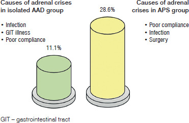
Importantly, adrenal crisis at presentation (or prior to diagnosis) tended to be more frequent in the APS cohort: 8 of 28 APS patients (28.6%) had at least one adrenal crisis, compared to 3 of 27 (11.1%) in the isolated AAD group. Although this marked difference (almost threefold) did not achieve statistical significance in our sample (p = 0.18), it may warrant further investigation in larger cohorts. All observed adrenal crises were precipitated by either a concurrent infection (e.g. pneumonia or gastroenteritis) or severe stress and were characterized by hypotension, hyponatraemia, hyperkalaemia, and/or hypoglycaemia requiring emergency hydrocortisone therapy.
Poor adherence to maintenance glucocorticoid treatment and intercurrent infections were the most prominent triggers of adrenal crises in both groups. In the APS group (yellow bar), 8 patients (28.6%) experienced at least one adrenal crisis, most often precipitated by missed medication doses or infections; in the AAD group (green bar), 3 patients (11.1%) had an adrenal crisis, with a similar distribution of precipitating causes. By contrast, major surgery and GI upsets (e.g. persistent vomiting leading to inability to take oral steroids) were less common precipitating factors – see Figure 1. These observations underscore the critical importance of patient and family education for treatment compliance and stress-dose steroid management to prevent life-threatening crises.
Laboratory findings
Laboratory evaluation at diagnosis revealed that ACTH levels were markedly elevated in virtually all patients of both groups, reflecting primary adrenal failure. In fact, 100% of tested individuals in both the AAD and APS groups had an ACTH level above the upper limit of the assay reference range (often well above 200 pg/ml), so this did not distinguish the groups (Table III).
Table III
Comparison of laboratory findings between isolated autoimmune Addison’s disease (AAD) and autoimmune polyendocrine syndrome (APS) groups at presentation (N = 55)
Both groups also showed low morning serum cortisol levels consistent with adrenal insufficiency (most had cortisol < 5 μg/dl at diagnosis). A slightly higher proportion of APS patients had documented low cortisol (82.1% vs. 74.1% in AAD), but this was not statistically significant (p = 0.70). In practice, any patient with clinical features suggestive of Addison’s and high ACTH was managed as having adrenal insufficiency regardless of the exact cortisol value, which might explain some “normal” cortisol values in the context of partial treatment or diurnal variation.
Significant differences emerged in the electrolyte and glucose abnormalities between the groups. Hyperkalaemia (elevated serum potassium) and hyponatraemia (low sodium) are hallmarks of mineralocorticoid deficiency. Hyperkalaemia was present in 74.1% of children with isolated AAD at diagnosis but in only 35.7% of those with APS (p = 0.009). Similarly, hypoglycaemia at presentation was far more frequent in the AAD group (88.9% of patients had blood glucose < 60 mg/dl on initial labs) compared to 28.6% of APS patients (p < 0.0001). These findings indicate more severe cortisol and aldosterone deficiencies in the isolated AAD patients. Hyponatraemia was also common, seen in 74.1% of AAD vs. 50.0% of APS patients, although the difference did not reach significance (p = 0.12). The high incidence of hyponatraemia in both groups likely reflects the fact that almost all patients, whether with isolated AAD or APS, presented with features of Addison’s disease sufficient for diagnosis.
In summary, isolated AAD patients tended to have more pronounced metabolic derangements at diagnosis than APS patients, possibly suggesting that APS patients’ adrenal insufficiency was recognized earlier or that they retained partial adrenal function longer. These laboratory disparities mirror the clinical observations of more frequent hypoglycaemic episodes and more severe dehydration in the AAD group.
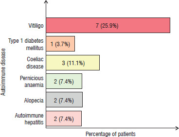
Among the 27 children in the isolated AAD group, we observed that a subset had other coexisting autoimmune disorders despite not qualifying as APS by definition. In isolated AAD patients, any additional autoimmune condition tended to be non-endocrine in nature (since the presence of another endocrine autoimmune disease would usually shift the diagnosis to APS-2). Vitiligo, an autoimmune skin depigmentation disorder, was the most common coexisting condition, found in 7 of 27 (25.9%) isolated AAD patients. Coeliac disease (autoimmune gluten sensitivity) was the next most frequent, present in 3 patients (11.1%). Other documented associations in the AAD cohort included alopecia areata (autoimmune hair loss) in 2 patients, pernicious anaemia in 2 patient, and autoimmune hepatitis in 2 patients – see Figure 2.
Figure 2
Associated autoimmune disorders among patients with isolated autoimmune Addison’s disease (n = 27)
Notably, none of the isolated AAD patients had coexisting type 1 diabetes or autoimmune thyroid disease – the presence of those would have met criteria for APS-2. This supports the classification of our “isolated” AAD group as lacking additional major endocrine autoimmunity. Nonetheless, the presence of vitiligo, coeliac disease, and other minor autoimmune conditions suggests an underlying predisposition to autoimmunity even in ostensibly isolated AAD.
In the APS group, by definition all patients had at least one other significant autoimmune condition (most commonly hypoparathyroidism and mucocutaneous candidiasis for APS-1, or type 1 diabetes and autoimmune thyroiditis for APS-2). Detailing these APS-defining conditions, in APS-1 patients, chronic mucocutaneous candidiasis and hypoparathyroidism were universally present alongside Addison’s disease, and some also had autoimmune hepatitis or alopecia. In APS-2 patients, autoimmune thyroiditis was present in all three, and one had type 1 diabetes as well.
Bars show the number of patients with each coexisting autoimmune condition, with percentages of the AAD group indicated. Vitiligo (skin depigmentation) was the most common associated disorder, affecting 25.9% of isolated AAD patients. This was followed by coeliac disease in 11.1%. Less frequent associations included alopecia areata (7.4%), pernicious anaemia (7.4%), and autoimmune hepatitis (7.4%). These findings demonstrate that even in children classified as having “isolated” AAD (no additional endocrine autoimmune diseases), there is a considerable overlap with other non-endocrine autoimmune conditions. In particular, cutaneous and GI autoimmune manifestations were relatively frequent, suggesting a need for periodic screening for these conditions during follow-up of paediatric Addison’s disease.
Discussion
Differentiating between isolated AAD and APS in children is an important clinical challenge. Although Addison’s disease is the common component, the broader context of autoimmunity differs substantially between isolated AAD and APS, carrying implications for monitoring and long-term management [14–16]. Our 15-year single-centre experience highlights several distinguishing features that can aid clinicians in anticipating complications and tailoring care for these rare patients.
In our cohort, familial autoimmunity was much more prevalent among APS patients. This aligns with existing literature indicating that APS (particularly APS-2) has a strong genetic and familial component, often clustering with other autoimmune diseases in families [17, 18]. A positive family history in a child with adrenal insufficiency should thus raise suspicion for an APS variant and prompt evaluation for other autoimmune diseases.
Children with isolated AAD tended to present with classic and pronounced features of cortisol and aldosterone deficiency. Significantly higher rates of hyperpigmentation and weight loss were observed among the AAD group. Hyper- pigmentation results from chronically elevated ACTH stimulating melanocortin receptors; its prominence in isolated AAD suggests a longer period of unmitigated ACTH elevation or more complete adrenal cortical destruction at presentation [19, 20]. Interestingly, vitiligo was the most common non-endocrine autoimmune comorbidity in isolated AAD patients, which is consistent with other reports linking AAD with cutaneous autoimmunity [21, 22]. The coexistence of vitiligo points to a generalized dysregulation of immune tolerance affecting both the adrenal cortex and melanocytes [23].
Weight loss in Addison’s disease is multifactorial, stemming from anorexia, GI malabsorption, hypercatabolism, and chronic dehydration due to aldosterone deficiency. Significantly higher weight loss in AAD patients likely reflects more severe hormonal deficits. In contrast, GI symptoms were more prominent in APS patients. Many APS-1 patients experience chronic mucocutaneous candidiasis and GI autoimmunity (such as autoimmune enteropathy or pernicious anaemia) [24, 25], which can cause abdominal symptoms independent of adrenal insufficiency. Thus, an Addison’s patient with disproportionate GI issues should be evaluated for APS-related gut pathology (e.g. malabsorption due to coeliac disease or chronic diarrhoea due to candidiasis).
A major finding was the difference in metabolic disturbances between the groups. Isolated AAD patients had dramatically higher hypoglycaemia and hyperkalaemia on initial presentation. Severe hypoglycaemia occurs in primary adrenal insufficiency due to cortisol deficiency impairing gluconeogenesis and increasing insulin sensitivity. Nearly 90% of isolated AAD patients had documented hypoglycaemia (vs. ~29% in APS), which may indicate that APS patients either retain partial endogenous cortisol production for longer or that their Addison’s disease was recognized and treated earlier in its course.
Similarly, hyperkalaemia indicates profound aldosterone loss; its significantly low incidence in APS could imply some residual zona glomerulosa function in those patients. Some APS patients (especially APS-2) might develop adrenal failure more insidiously or have an overlap with genetic syndromes that modulate disease expression, resulting in milder electrolyte abnormalities early on.
The possibility of residual adrenal function in APS patients is intriguing. Recent studies have reported that a subset of AAD patients maintain intermittent or partial adrenal steroid production, raising questions about differences in disease pathogenesis [26]. One hypothesis is that the polyendocrine context in APS might allow periods of immune remission or contain autoimmunity that spares some adrenal tissue. Alternatively, there may be adrenal stem cell niches capable of regenerating cortical cells in some patients [27]. These concepts are supported by reports of APS patients who have recovered some adrenal function years after diagnosis [28]. Our findings of milder biochemical abnormalities in autoimmune polyglandular syndromes APS align with the notion that total adrenal destruction might be less uniform in those patients, at least at presentation. This could inform future therapies aimed at preserving adrenal tissue [29].
Emerging treatments are exploring strategies to preserve residual adrenal function or limit autoimmune-mediated adrenal damage. For example, administration of long-acting ACTH analogues (tetracosactide) has been shown to stimulate adrenal steroidogenesis and potentially delay the complete loss of function in AAD [30]. Additionally, immunomodulatory therapies such as rituximab combined with depot ACTH have been trialled to prolong adrenal function by targeting the autoimmune process [31]. These experimental approaches (referenced by Smans et al. [25] and Baxter [26] et al.) have shown some promise in improving endogenous cortisol production in select patients. Our observation that APS patients may have partial function for longer suggests that they could especially benefit from such interventions, if identified early.
Another noteworthy observation was the rate of adrenal crises. We observed a higher frequency of APS patients experiencing adrenal crises. Although not significant, the difference is in line with larger studies. For instance, a German registry reported that APS patients had over 2.5-fold higher risk of adrenal crisis compared to those with isolated AAD [32].
APS patients may be more susceptible to crises due to the presence of additional stressors (e.g. intercurrent illnesses from other autoimmune conditions) and perhaps the complexity of managing multiple diseases. Our data also reaffirmed known precipitating factors for crisis such as poor compliance with daily steroid therapy and delayed stress dosing during infections [33]. These are modifiable factors – underscoring that patient education and adherence are lifesaving priorities in chronic adrenal insufficiency management.
From a clinical perspective, our findings highlight the need for a tailored approach to management for these patients. Children with isolated AAD, given their propensity for severe metabolic disturbances, should be closely monitored for hypoglycaemia and hyperkalaemia, especially at diagnosis and during intercurrent illnesses.
Aggressive patient education on “sick day” rules is essential. Meanwhile, for APS patients, clinicians should maintain a high index of suspicion for other autoimmune complications – for example, screening for coeliac disease, thyroid dysfunction, or diabetes at regular intervals. Anticipatory guidance for APS patients might also involve closer follow-up and perhaps a lower threshold for hospitalization during illnesses, considering their observed higher (albeit not significant here) crisis risk.
This study is limited by its retrospective design and small sample size inherent to a single-centre experience with rare disorders. The statistical power to detect differences, especially in outcomes such as adrenal crisis, is limited. Additionally, APS itself is heterogeneous; combining APS-1 and APS-2 patients under one umbrella is a simplification, though necessary due to small numbers. Nonetheless, our 15-year experience provides valuable insights into the paediatric phenotype of Addison’s disease in Iraq and contributes data from a region where such information is sparse. The consistency of our observations with reports from other settings (for example, the prominence of hyperpigmentation and vitiligo in isolated AAD, or the increased familial autoimmunity in APS) lends credibility to our findings and suggests that they may be generalizable, while also highlighting region-specific needs such as early diagnosis to prevent crises [34].
Looking ahead, future research should focus on prospective, multicentre studies to better define the risk factors for adrenal crisis and other outcomes in these patients. Genetic analysis (for example, of the AIRE gene in APS-1 or human leukocyte antigen associations in APS-2) in APS-2) could further elucidate why some patients develop isolated AAD vs. APS [35].
Moreover, trials of immunotherapies or adrenal-preserving treatments would be greatly beneficial. As our understanding of the autoimmune attack on the adrenal gland improves, there is hope that targeted interventions might prolong the functional life of the adrenal cortex in newly diagnosed Addison’s disease patients.
The practical value of this work lies in improving early recognition, tailored screening, crisis prevention, and individualized management of paediatric AAD by clearly distinguishing isolated AAD from APS at the bedside. Other than the diagnostic value, these findings support the consideration of a more individualized management approach that integrates environmental and lifestyle factors, including nutritional optimization, physical activity counselling, and emerging strategies such as modulation of the gut microbiota (e.g., probiotics), as adjunctive measures aimed at supporting metabolic stability, immune regulation, and long-term outcomes in affected children [9, 36].
Conclusions
In conclusion, this study demonstrates that distinguishing isolated AAD from APS in paediatric patients is clinically important and feasible based on characteristic features. Children with isolated AAD were more likely to have classic manifestations of adrenal insufficiency – notably severe hyperpigmentation, weight loss, hypoglycaemia, and electrolyte disturbances – whereas those with APS often had a positive family history and tended to suffer adrenal crises more frequently (though not significantly so in our sample). Both groups demand vigilant management, but APS patients require broader surveillance for multiple autoimmune diseases and careful crisis prevention strategies. Ultimately, recognizing whether a child’s Addison’s disease is isolated or part of APS informs prognosis and guides a more individualized treatment plan. Emphasis on patient education (particularly regarding adherence to steroid replacement and stress dosing during illness) and infection prevention is critical in all cases to reduce the risk of adrenal crises. Our 15-year experience adds to the growing body of evidence that early, tailored interventions can improve outcomes for children with these rare yet challenging endocrine disorders. Both isolated AAD and APS-associated Addison’s disease are linked to an increased risk of other autoimmune diseases during the patient’s lifetime, highlighting the importance of long-term clinical follow-up.