


Meckel’s diverticulum (MD) is the most common congenital anomaly of the gastrointestinal tract, and according to several studies its prevalence is about 0.6–4% [1]. The exact location of MD is mentioned in the literature and in textbooks, and its characteristics usually follow the rule of twos: 2% of the general population, 2 feet proximal to the ileocecal valve, 2 inches in length, and 2 : 1 male: female ratio [2].
A 42-year-old patient presented with a 24-hour history of sudden-onset abdominal pain, nausea without vomiting, and inability to pass flatus. His medical history was unremarkable for any conditions, and he had not undergone any previous abdominal surgery in the past. He denied any previous similar symptoms before the index episode with no fevers, bleeding per rectum, weight loss, or any other symptoms pointing in the direction of underlying malignancy or inflammatory bowel disease. In addition, his family history was unremarkable for such conditions. Upon physical examination, he was noted to have abdominal distention, with hyperactive bowel sounds and tenderness on palpation on all 4 quadrants with signs of localised peritonism, especially in the middle right side. On laboratory findings, marked leukocytosis and elevated C-reactive protein were noted. All other lab tests including liver function tests and amylase were normal. The patient underwent a computed tomography (CT) scan on an emergency basis to investigate the cause of his symptoms. CT scan revealed a fluid-filled blind-ending loop around the terminal ileum with fat stranding and dilated loops of small bowel proximal to that. The appendix was noted to be normal, and no overt masses were noted (Figures 1, 2). Based on the diagnosis of complicated small bowel obstruction in a virgin abdomen, he was taken to theatre for an emergency laparotomy. Upon surgical exploration, he was found to have an inflamed Meckel’s diverticulum with relative sparing of its base with a persistent band from the diverticulum to the small bowel mesentery (mesodiverticular band). Loops of small bowel were found to be twisted around the inflamed diverticulum and the mesodiverticular band, which acted as a lead point for “knotting of the small bowel”. The mesodiverticular band was carefully ligated and divided. The trapped loops of the small bowel were reduced and did not present any signs of ischaemia. The Meckel’s diverticulum was noted to have a long axis with a relatively narrow base. A decision was made to perform diverticulectomy rather than a formal small bowel resection, and therefore a linear stapler was fired across the anti-mesenteric border of the small bowel at the base of the diverticulum. The staple line was further inverted with interrupted seromuscular absorbable sutures. The postoperative course was unremarkable, with a return of bowel function as early as postoperative day 1 and rapid resolution of the ileus. The patient was safely discharged back to the community after 3 days and was followed up again 2 weeks later in the outpatient clinic with no problems encountered. Final pathology revealed Meckel’s diverticulum without any presence of ectopic tissue with dimensions 6.7 cm in length and 2.9 cm in width. No evidence of malignancy was noted.
Figure 1
Axial images of the preoperative CT scan showing a blind-ended fluid-filled loop in the area of the terminal ileum
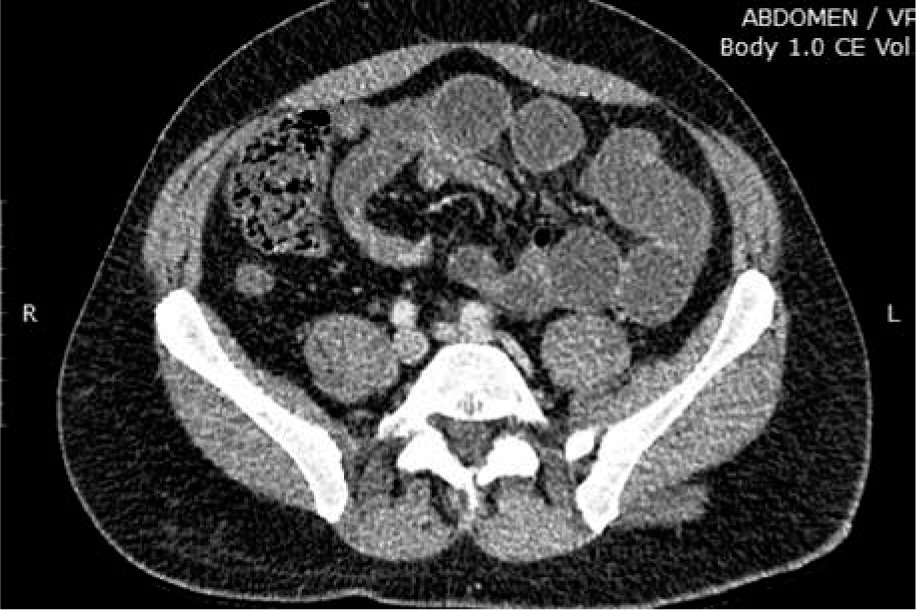
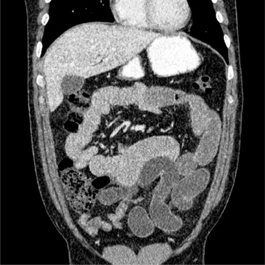
Figure 2
Coronal CT scan showing signs of internal herniation in the area of the terminal ileum with dilatation of small bowel loops and a normal- looking appendix
While the majority of MDs remain asymptomatic, they can also present in a complicated fashion, most commonly with bleeding, obstruction, and inflammation/perforation. In adults, diverticulitis and small bowel obstruction represent the most common presentations according to a study from Parvanescu et al., with the authors describing the age of 40 years as a cutoff point. Before the age of 40 years, bleeding is the most common symptomatic presentation, while after 40 years of age, diverticulitis and small bowel obstruction ensue at a higher degree [3]. When MD causes small obstruction, the most common mechanisms are intussusception, protrusion of the diverticulum into the small bowel lumen, trapped enteroliths, diverticulitis, volvulus around a remaining mesodiverticular band causing internal herniation, and in extreme cases “knotting of the small bowel” or on rare occasions entrapment in a hernia sac, called Littre’s hernia [4–7]. MD can also harbour malignancy at an incidence much greater than other sites in the terminal ileum [8].
A report from Mayo clinic encompassing 52 years of experience and 1476 patients in total, which consists of the largest single-centre case reported to date, reports a 16% rate of Meckel’s diverticula being symptomatic [9]. While the authors neither support nor reject resection of an incidentally found MD, they recommend a selective approach for resection in patients with any one of the aforementioned features. A systematic review by Zani et al. reported that the number of MD resections per 100,000 population decreased significantly after paediatric age, and they also reported a higher complication rate of resecting versus leaving the MD in situ, with a number needed to treat to prevent 1 death from MD calculated at 758, thus advocating against resection of an incidentally found MD [10]. Regarding the decision for diverticulectomy versus formal small bowel resection for symptomatic MD, the decision varies according to the indication as well as the morphology of the diverticulum. A study by Varcoe et al. identified the height-to-diameter ratio of the diverticulum with a cutoff of 2 as a marker of the presence of ectopic tissue within the tip of an MD versus within the whole MD (as is the case according to the authors for diverticula with a ratio less than 2) [11]. Based on these findings, they advocate formal resection for short diverticula, stating also that both the external appearance and palpation cannot reliably inform correct decisions. Apart from the presence of ectopic mucosa, the integrity of the base (healthy versus non-healthy), the state of adjacent ileum as well as the presence of tumour inside an MD also guides decisions, with diverticulectomy being probably a sound approach for diverticulitis with a non-compromised base while base involvement by inflammation, perforation, presence of tumour, as well as resecting for bleeding mandate segmental or wedge bowel resection [12].
With regards to small bowel obstruction in a virgin abdomen, the classic surgical dogma has been in favour of aggressive and early surgical management, with the most common cause of obstruction being adhesions (49%) followed by malignancy (10%) and then others (24%), while 17% of patients remain without a diagnosis after laparotomy, according to a recent meta-analysis [13]. Non-operative management appears to be an option for clinically stable patients who are closely monitored, without concerning signs on CT, and without evidence of a closed-loop obstruction [13–15]. It is, thus, reasonable to conclude that Meckel’s diverticulitis causing small bowel obstruction in a virgin abdomen presents an extremely rare entity because it is a rare entity per se belonging to the subgroup of least common causes of small obstruction in a virgin abdomen, thus forming an unusual combination of 2 different entities, which, to our knowledge, is the first explicitly reported in the literature. With regards to the means of pre-operatively establishing such a diagnosis of complicated Meckel’s diverticulum in the setting of small bowel obstruction (in which the modality of imaging of choice in the emergency setting is the CT scan), the available literature only identifies as potential findings of an MD on a CT scan a fluid or gas-filled blind-ending loop, which is, however, difficult to decipher from small bowel loops [16]. Signs of inflammation, with thickening of the wall of this blind-ending tubular structure, fluid/abscess, and fat stranding in the vicinity, especially in the presence of an identified normal appendix, have been reported as signs of Meckel’s diverticulitis on CT [17]. Laparoscopic surgery has been performed for small bowel obstruction, showing better postoperative short-term outcomes than open surgery [18].
In the aforementioned case, it is evident that the treatment algorithm was guided by the mode of presentation as well as by the morphology of the MD, thus leading to diverticulectomy versus formal small bowel resection. Available data on decision-making regarding the management of complicated small bowel obstruction in a virgin abdomen and CT signs suggestive of Meckel’s diverticulitis have been reaffirmed by our case presentation.
Small bowel obstruction in a virgin abdomen with signs of peritonitis and closed-loop obstruction presents a true surgical emergency and should be aggressively treated. Mesodiverticular band is a rare cause of small bowel obstruction in Meckel’s diverticulum in adults. The decision to perform diverticulectomy versus formal small bowel resection should be guided by the indication for which Meckel’s is operated on (bleeding versus obstruction versus tumour versus diverticulitis) and the morphology of MD per se. Especially in cases of obstruction, apart from the usual scenarios of intussusception, inversion, or trapped enteroliths, the presence of a mesodiverticular band or a fibrous cord from the diverticulum to the umbilicus (persistent vitelline duct) causing internal herniation of the small bowel in conjunction with diverticulitis of the MD should be in the diagnostic arsenal of the acute care surgeon. Regarding asymptomatic Meckel’s diverticula, these usually remain asymptomatic and thus do not warrant treatment if incidentally found, unless they have worrisome features such as presentation in young adults, in male patients, with length greater than 2 cm, or presence of ectopic mucosa, according to most reports to date. However, more data on this rare entity are needed to further inform our decisions.