Introduction
In recent years, metabolic diseases began to present a significant global health problem in the pediatric population. According to the World Health Organization (WHO) fact sheet [1], over 390 million children and adolescents aged 5–19 years were overweight in 2022, including 160 million who were living with obesity. The United Nations Children’s Fund (UNICEF) reported that between 2000 and 2022, the percentage of obese children aged 5–19 increased from 3% to 9.4% [2]. Furthermore, the population of obese children under the age of 5 increased to 35 million [1]. Additionally, boys were significantly more likely to be obese than girls [3]. Obesity is strongly associated with the occurrence of complications, primarily metabolic dysfunction–associated steatotic liver disease, insulin resistance, diabetes mellitus, and metabolic syndrome [4].
The incidence of pediatric diabetes has increased by 39.4%, with the greatest rise observed in low-income countries [5]. The pathophysiology of pediatric obesity is multifactorial. The main causes remain an unhealthy diet and low physical activity, as modifiable environmental factors. An excessive intake of high-calorie, nutrient-poor foods, and sugar-sweetened beverages, combined with a sedentary lifestyle, is strongly associated with an increased risk of obesity in the pediatric population [6]. It also encompasses behavioral factors, including poor sleep quality and the resulting chronic inflammatory state [7]; neuroendocrine factors, including the regulation of hunger and satiety associated with disturbances in the secretion of leptin, ghrelin, peptide YY (PYY), glucagon-like peptide-1 (GLP-1), and insulin [8]; genetic factors, including single-nucleotide polymorphisms, copy number variations, as well as insertions and deletions [9].
In recent years, there has been a marked increase in interest in the gut microbiota and the impact of its dysbiosis on the development and progression of metabolic diseases in children. The gut microbiota has been shown to comprise approximately 1014 microorganisms, including bacteria, viruses, fungi, and archaea, predominantly colonizing the large intestine. This corresponds to a number approximately 10 times higher than the total number of human somatic cells and encodes about 100 times more genes than the human genome [10]. It demonstrates that an appropriate composition of the gut microbiota enhances intestinal epithelial barrier integrity, thus protecting against the translocation of pathogens originating from ingested or inhaled sources [11]. The aim of this review is to describe and analyze the current state of knowledge on the impact of the gut microbiota on the development and progression of metabolic diseases in children, with particular emphasis on pathophysiological mechanisms and potential therapeutic options.
Gut microbiota
The gut microbiota is described by many scientists as a superorganism. This is primarily due to the enormous number of microorganisms that comprise it and the diverse functions it performs to maintain the body’s homeostasis. Colonization of the gut microbiota begins at birth, which is also reflected in placental tissues [12]. The mode of delivery also influences the initial composition of the neonatal microbiota. Cesarean delivery deprives the neonate of colonization by maternal fecal microbiota, and species of the genus Bacteroides, predominantly present in infants delivered vaginally, are replaced by Clostridium species [13]. The composition of the intestinal microflora is significantly influenced by infant feeding practices, especially during the first year of life. Breast milk is a source of Bifidobacterium bacteria (mainly B. longum and B. breve), as well as Lactobacillus, Bacteroides, and Streptococcus, which use human milk oligosaccharides as metabolic substrates [14]. Breast milk also provides immunoglobulin A (IgA) and anti-inflammatory factors, and additionally lowers the pH of the intestines, which protects against colonization by pathogenic microorganisms. Breastfed infants have reduced numbers of Enterobacteriaceae and very low concentrations of species such as Clostridium difficile, Escherichia coli, and Staphylococcus [15]. In contrast, formula feeding is associated with greater diversity of bacteria of the genera Bacteroides, Enterococcus, Staphylococcus, Clostridium, and Enterobacteriaceae, while colonization by Bifidobacterium is delayed [14]. In addition, the introduction of cow’s milk causes an increase in the number of Bacillota (formerly Firmicutes) bacteria and bacteria with pro-inflammatory properties [16]. Therefore, it is recommended that the optimal breastfeeding period should be at least 6 months (WHO, 2023). It has been demonstrated that the use of antibiotics also disrupts the structure of the gut microbiota. In a cohort study involving children aged 2–7 years, macrolide use was associated with a persistent decrease in Actinobacteria and an increase in Bacteroidetes and Proteobacteria [17]. The microbiota mainly affects the integrity and development of the intestinal epithelium, regulates immune responses, but also has anti-cancer properties. Importantly, its composition begins to take shape during childbirth. It has been shown that the microflora of children born vaginally contains up to 72% of the mother’s fecal microflora, while in children born by cesarean section, this percentage is almost half as much [11]. The diversity of Escherichia, Bifidobacterium, and Bacteroides bacteria is significantly greater in newborns delivered vaginally, while Pseudomonas, Lactobacillus, and Acinetobacter bacteria dominate in infants delivered by caesarean section. In addition, significantly higher levels of IgA and immunoglobulin M are observed in the umbilical cord blood of newborns delivered vaginally, which is attributed to increased pressure in the fetal-placental vessels during uterine contractions during labor [18]. The timing of birth also appears to be important: preterm infants have significantly higher levels of Enterobacter, Staphylococcus, and Enterococcus species, which are associated with increased morbidity and mortality in children [19]. The microbiome changes with age. By 18 months of age, the abundance of Enterobacteriaceae, Bifidobacteriaceae, and Clostridiaceae decreases by approximately 25-, 5-, and 3.5-fold, respectively, compared with levels observed in newborns [20]. At around 3 years old, a child’s microbiome is similar to that of an adult [19]. Some studies indicate that living in rural areas is also associated with greater diversity in a child’s microbiome. Intestinal bacteria can be broadly divided into three main enterotypes: Bacteroides, Prevotella, and Ruminococcus. The role of this intestinal microflora focuses mainly on the fermentation of polysaccharides. Bacteria, including those from the Bacillota and Bacteroidota phyla, break them down into short-chain fatty acids (SCFAs), primarily acetate, butyrate, and propionate. These metabolites play important roles in host metabolism, with butyrate serving as a major energy source for intestinal epithelial cells [21]. In addition, acetate serves as a substrate in lipogenesis and gluconeo-genesis. Butyrate supports anti-inflammatory effects by inhibiting histone deacetylase activity [22]. SCFAs lower the pH of the intestinal lumen and reduce oxygen availability, thereby protecting commensal strains and inhibiting the growth of pathogenic Salmonella Typhimurium, Clostridium difficile, and Escherichia coli. The integrity of the intestinal barrier also depends on other factors. One of these is the intestinal mucus layer, which serves as a protective barrier between the gut microbiota and the intestinal epithelium. Impaired mucus production may promote colonization by pathogenic organisms such as Citrobacter rodentium and Salmonella Typhimurium, increasing the risk of intestinal injury. In addition, the microflora produces signaling molecules (MAMPs) such as lipopolysaccharide (LPS), which, when bound to toll-like receptor (TLR) and nucleotide-binding oligomerization domain receptors, activate macrophages to produce pro-inflammatory cytokines interleukin-1 (IL-1), interleukin-6 (IL-6), and interleukin-23, affecting the balance between T helper 17 cells and regulatory T cells and strengthening the epithelial barrier [23].
Role of the gut microbiota in obesity
Overweight and obesity are a growing problem in the pediatric population. In 2015, over 100 million children worldwide were estimated to have obesity [24]. Data from 2025 indicated that this number had almost doubled, to approximately 188 million. Obesity is diagnosed in children who exceed the 95th percentile of weight for their age and gender in centile charts, and overweight in those who exceed the 85th percentile. In children under 5 years of age, a body mass index greater than 2 standard deviations is calculated [25]. Obesity is a disease that usually results from a variety of causes, primarily an unbalanced diet and an inactive lifestyle, but also endocrine causes such as hypothyroidism, Cushing’s syndrome, growth hormone deficiency, or genetic factors in disorders of the lectin/melanocortin pathway, Prader-Willi syndrome and Beckwith-Wiedemann syndrome [26]. In recent years, many studies have suggested that an important factor in childhood obesity may be an imbalance in the composition of the gut microbiota. The diversity of the gut microbiota in children can depend on age, stage of puberty, ethnic group and gender. The microbiome varies significantly with age [27], and its diversity increases until adulthood [28, 29]. As children grow older, a general decline in the abundance of Bacteroides dorei, Parasutterella excrementihominis, Clostridium clostridioforme, and Faecalibacterium prausnitzii has been observed [27]. Significant changes in microbiota diversity occur during the first year of life. Until the age of 6 months, Lactobacillus, Bifidobacteria, and Bacteroides dominate, while Clostridium, Bacillus, Roseburia, and Faecalibacterium also appear. In the 7–12-month age group, Providencia, Akkermansia, Acinetobacter, and Veillonella dominate, while in the 13–24-month age group, Comamonas, Clostridium, and Novibacillus are predominant [28, 30]. The distribution of the most common gut bacteria according to age is presented in Table I. The gradual transition from liquid to solid foods is associated with a decrease in the abundance of Bifidobacterium and an increase in that of Bacillota. Importantly, higher abundance of Coriobacteriaceae, Erysipelotrichaceae, Lachnospiraceae, and Ruminococcaceae, and lower abundance of Verrucomicrobia and Atopobium parvulum at 3–4 months of age were observed in infants who became overweight as early as 12 months of age [31]. Around the age of 5, the composition of the microbiome begins to stabilize, with Bacteroides, Prevotella, and Bifidobacterium predominating in healthy children [31]. At this time, the diversity of the gut microbiota plays an important role in immune functions. Bacteria that are constantly present from birth until the age of 5 include Bifidobacteria, Lactobacilli, Providencia, Bacteroides, Akkermansia, Faecalibacterium, Streptococcus, Collinsella, Anaerobutyricum, and Prevotella [28]. In overweight school-aged children, differences are observed, with a higher number of Clostridium and lower numbers of Akkermansia muciniphila, Oscillospira, and Rikenellaceae. In adolescents, changes in the abundance of Bifidobacterium and Clostridium are observed compared to adults; in obese children, an increase in the abundance of Actinomyces is also observed [31]. Studies show that the gut microbiome also influences sex hormone levels, which may be important for growth during this period [29]. It has been shown that changes in the composition of a child’s gut microbiota can vary by ethnic group as early as 3 months of age and persist throughout childhood. Interestingly, approximately one-third of the taxa overlap with differences observed in adults [32]. According to studies, African American children are more likely to become obese than European children. In the general population, they have been shown to have increased abundance of Ruminococcaceae, Anaerotruncus, Marvinbryantia, Oxalobacter, Prevotella, Senegalimassilia, and Slackia compared to the European population. In the group of obese African American children, Klebsiella, Megasphaera, and Coriobacteriaceae predominate [33] compared to children of normal weight. Another study in Singapore involving infants from three ethnic groups showed that Indian infants exhibit a predominance of Bifidobacterium and Lactobacillus, Chinese infants present a huge number of Bacteroides and Parabacteroides, while the composition of the microbiota in Malay infants is intermediate between these two groups [34]. In a study conducted in the United States, higher abundances of Ruminococcus gnavus, Collinsella, and Sutterella were found in Nigerian infants [35], whereas in children of Chinese ethnicity between the ages of 0 and 36 months, higher abundances of Bifidobacteriaceae, Lachnospiraceae, Ruminococcaceae, and Faecalibacterium were observed [36]. Changes are also observed depending on the child’s gender. As early as the first year of life, higher abundances of Alistipes, Anaeroglobus, Bacillota, and Proteobacteria were observed in boys [37]. In older children, higher levels of Akkermansia and Bifidobacterium were observed in girls, while boys showed increased levels of Prevotella and Escherichia [38]. The composition of the gut microbiome in obese children differs significantly from that of healthy, slim individuals and shows reduced diversity. The species that displace the normal gut microbiota are mainly Bacteroides plebeius, Parasutterella excrementihominis, Parabacteroides distasonis, Bilophila wadsworthia, Clostridium symbiosum, Megamonas funiformis, Allisonella histaminiformans, Prevotella stercorea, and Oxalobacter formigenes [39]. These microbes are responsible for the increased ability to ferment undigested polysaccharides into SCFAs, which constitute a larger pool of substrates for lipogenesis in the liver and triglyceride accumulation in adipocytes. SCFA activate G protein-coupled receptors located on enterocytes and adipocytes. This increases insulin sensitivity through an increase in GLP-1, which in chronic overproduction leads to adipose tissue growth [40]. The displacement of Bifidobacterium and Bacteroides, combined with the proliferation of Bacillota and Proteobacteria, is associated with satiety disorders due to elevated ghrelin levels and reduced leptin sensitivity. In addition, intestinal dysbiosis leads to increased intestinal permeability, release of lipopolysaccharide into the bloodstream, and disruption of the hypothalamic–pituitary–adrenal axis, leading to hypercortisolemia [41]. Disturbed gut microbiota also disrupts tryptophan metabolism, leading to decreased serotonin, and its reduced levels contribute to negative mood and increase appetite under the influence of emotions [42]. A schematic overview of the relationship between gut microbiota, intestinal barrier dysfunction, inflammation, and metabolic diseases in the pediatric population is presented in Figure 1.
Figure 1
The association between gut microbiota and the prevalence of metabolic diseases in the pediatric population. An increase in the abundance of dysbiotic bacteria in children is caused by various factors (1), leading to impaired intestinal epithelial function (2), and consequently to pancreatic impairment by macrophages, dendritic cells, and T lymphocytes (3). Increased intestinal permeability leads to elevated low-density lipoprotein cholesterol levels, and consequently to the accumulation of white adipose tissue and the development of obesity (4). There are also protective factors that promote the growth of symbiotic bacteria (5), which support the proper functioning of the intestinal epithelium (6)
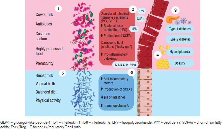
Table I
Representative gut microbiota taxa associated with eubiosis and dysbiosis across different stages of childhood
Obesity complications
Type 2 diabetes
Type 2 diabetes (T2D) is a disease primarily resulting from impaired insulin sensitivity in body cells. Intestinal dysbiosis may be one of the factors linking obesity to the occurrence of T2D, but it may also be a direct cause of this disease, regardless of the coexistence of obesity [43]. Disruption of the commensal composition of the intestinal microbiome leads to increased intestinal permeability and, consequently, metabolic endotoxemia resulting from the entry of pathogens and pro-inflammatory factors into the systemic circulation. A significant role is attributed to LPS, which is a component of the wall of Gram-negative bacteria. It activates TLR4 and TLR2 receptors on monocytes, which induce an inflammatory response and impair the function of pancreatic beta cells responsible for insulin secretion [44]. In a group of subjects aged 7–17 years, a significant association was observed between T2D, obesity, and gut microbiota composition. The study focused primarily on the genera Prevotella, Dorea, Faecalibacterium, and Oscillospira, which showed a positive correlation with the presence of abdominal obesity and T2D in this age group [45]. Another study reported lower species diversity of Bacillota and Proteobacteria in 10–16-year-old subjects with Homeostasis Model Assessment of Insulin Resistance (HOMA-IR) results indicative of insulin resistance [46]. Children with T2D have significantly lower concentrations of Faecalibacterium prausnitzii, Anaerostipes hadrus, and Roseburia intestinalis [43, 47], which are important for butyrate-producing bacteria. This SCFAs has been implicated in the regulation of insulin sensitivity and the maintenance of intestinal barrier integrity [47]. In addition, dysbiotic microorganisms activate the action of liver macrophages, which directly induce insulin resistance in hepatocytes by secreting the IGFBP7 protein. The association between obesity and the development of diabetes is mediated, in part, by excess adipose tissue. Adipocytes secrete the chemokine monocyte chemoattractant protein-1, which activates monocytes and T lymphocytes, thereby exacerbating inflammation and damaging pancreatic cells. In turn, activated natural killer and group 1 innate lymphoid cells secrete interferon-γ, which stimulates M1 macrophages with pro-inflammatory function. These macrophages release the inflammatory mediators tumor necrosis factor-α (TNF-α), IL-6, and IL-1β, which cause reduced sensitivity of fat, muscle, and liver tissue receptors to insulin [48]. The effect of antidiabetic drugs, including metformin and sulfonylurea, as well as statins, proton pump inhibitors, and non-steroidal anti-inflammatory drugs has also been studied. One study found no significant effect of these drugs on the composition of the gut microbiota [43]. Other studies indicate significant changes in the microbiome of people using drugs from these groups. Empagliflozin, a sodium–glucose cotransporter 2 inhibitor, increases the diversity of commensal species of Roseburia, Eubacterium, and Faecalibacterium and reduces the number of harmful species of Escherichia/Shigella, Bilophila, and Hungatella in patients with T2D. It has also been shown that the GLP-1 agonist liraglutide has a protective effect by increasing the abundance of Collinsella, Akkermansia, and Clostridium species and inhibiting the growth of Fusobacteria [47].
Hyperlipidemia
Hyperlipidemia is one of the primary metabolic disorders coexisting with obesity in the pediatric population. It is estimated that up to 40% of pediatric patients with obesity develop dyslipidemia (4). In diagnostic tests, hyperlipidemia in children aged 10–18 years can be diagnosed at low-density lipoprotein cholesterol (LDL-C) levels > 130 mg/dl, total cholesterol ≥ 200 mg/dl, and triglycerides > 130 mg/dl. Screening is important in the prevention of early atherosclerotic changes [49]. An increasing number of studies emphasize that the composition of the gut microbiota in the pediatric group has a multidirectional impact on the occurrence of hyperlipidemia associated with obesity [50]. Intestinal dysbiosis negatively affects lipid metabolism by regulating fat absorption, fatty acid synthesis, and digestive processes. It has been demonstrated that a reduced abundance of bacteria from the Lachnospiraceae and Ruminococcaceae families and the genera Akkermansia, Bacteroides, Roseburia, and Faecalibacterium is associated with hyperlipidemia in children. This results in decreased production of SCFAs, which play a crucial role in lipid metabolism regulation. SCFAs contribute to strengthening the intestinal barrier integrity, regulation of appetite through their influence on gut hormones, activating of receptors involved in the browning of white adipose tissue, and increasing thermogenesis [51]. A study in children aged 10–12 years demonstrated that the gut microbiota is also involved in bile acid metabolism, including processes such as deconjugation, epimerization, and 7α-dehydroxylation, leading to the formation of secondary bile acids. These compounds activate receptors that regulate cholesterol metabolism; for example, activation of the farnesoid X receptor reduces cholesterol utilization, resulting in excess cholesterol accumulation in the blood [50]. A study in children aged 8–14 years showed that a reduction in numbers of Christensenellaceae and Akkermansia promotes abnormal lipid metabolism and may lead to cardiometabolic complications in later years [52]. Another study in a 7–17-year-old group noted higher numbers of Faecalibacterium and Oscillospira in the presence of hyperglycemia. Prevotella, Dorea, Faecalibacterium, and Lactobacillus have a similar effect in correlation with triglyceride levels [45]. In a different group of children aged 11–15 years, overweight and obesity were negatively associated with the presence of the genera Vibrio and Salmonella. Although many studies are performed on relatively small cohorts (approximately 100 patients), it can be concluded that the presence of dysbiotic microbiota causes increased levels of total cholesterol, LDL-C, and triglycerides, which are characteristic features of hyperlipidemia in children [53].
Gut microbiota in T1D
For many years, type 1 diabetes (T1D) was regarded as a disease predominantly driven by genetic factors. Mutations in human leukocyte antigen (HLA) genes (HLA-DR3, HLA-DR4, and HLA-DQ8) play a major role, but in recent years, the importance of insulin, protein tyrosine phosphatase non-receptor type 22, and cytotoxic T-lymphocyte-associated protein 4 has also been confirmed [54]. In T1D, the immune system begins to produce autoreactive T lymphocytes (CD8+ cytotoxic T cells and CD4+ helper T cells) [55] and autoantibodies against pancreatic β-cells, including anti-glutamic acid decarboxylase 65, anti-insulinoma-associated antigen-2, anti-insulin antibodies, and anti-zinc transporter 8 [56]. In addition to genetic predisposition, environmental factors are believed to play a role in triggering this process; these include viral infections, e.g., enteroviruses (Coxsackie B), as well as diet-related factors [57]. In recent years, increasing evidence suggests that susceptibility to T1D in children is significantly influenced by the gut microbiota [58]. Comparative analyses of the gut microbiota revealed that children with T1D, compared with healthy controls, showed an increased relative abundance of Bacteroides dorei, Bacteroides vulgatus [59] and Bifidobacterium spp. and a reduced concentration of Streptococcus thermophilus [60]. These bacteria are involved in metabolic pathways related to the production of SCFAs, which play a key role in maintaining intestinal barrier integrity and gut homeostasis. Reduced production of SCFAs leads to increased intestinal permeability. It is also attributed to a diminished concentration of SCFA-producing bacteria, particularly Clostridium clusters IV and XIVa (including Faecalibacterium prausnitzii) and Roseburia, which are crucial for maintaining the integrity of the intestinal barrier [61]. Increased intestinal permeability (“leaky gut”) allows bacterial fragments and endotoxins (e.g. LPS) to penetrate the bloodstream and intestinal lymphatic tissue. This activates local and systemic immune responses and may promote autoimmunity against pancreatic β-cells [62]. This occurs through stimulation of dendritic cells and macrophages, which present antigens to T lymphocytes and produce proinflammatory cytokines, including IL-1β, IL-6, and TNFα [63]. In children with T1D, the presence of dysbiotic bacteria may influence the occurrence of obesity. According to one study, an increase in the number of Prevotella and Bacteroides increases the synthesis of branched-chain amino acids. This promotes insulin resistance, which, together with insulin therapy, leads to increased deposition of subcutaneous fat [64].
Prevention and treatment
The basis of childhood obesity treatment remains a well-balanced diet and physical activity. In the context of intestinal dysbiosis, increasing dietary fiber intake has been associated with increased numbers of Faecalibacterium prausnitzii and Lachnospira spp., Eubacterium, and Roseburia, which protect against obesity. Additionally, the consumption of complex polysaccharides, plant proteins, and unsaturated fats enriches the gut microbiota with species producing SCFAs [27], which contribute to the maintenance of intestinal barrier integrity and gut homeostasis. It is also recommended to reduce the consumption of simple sugars, which increases the number of dysbiotic bacteria: Bacteroides dorei, Turicibacter sanguinis, Bacteroides vulgatus, and Bacteroides fragilis [29].
Probiotics, prebiotics, and synbiotics have also been investigated as potential adjunctive interventions in the treatment of obese children with intestinal dysbiosis. Prebiotics are undigested food components that stimulate the growth of specific bacterial strains in the colon. The most commonly used prebiotics are fructans, inulin, fructooligosaccharides, and lactulose. They primarily increase the number of Bifidobacterium and Lactobacillus species, which are associated with beneficial effects on gut homeostasis and immune regulation [65]. Additionally, these bacteria increase GLP-1 and PYY and decrease ghrelin, which stabilizes appetite in pediatric patients. Furthermore, regulating the expression of the lipoprotein lipase inhibitor protein reduces lipid storage in adipose tissue [66]. Prebiotics provide the best treatment results. Prebiotic supplementation in children aged 7–12 years was found to reduce body weight by an average of 3.1% and body fat by an average of 2.4% over 16 weeks [67]. According to the WHO, probiotics are live microorganisms that, when administered in appropriate amounts, have a beneficial effect on the patient’s health. The most commonly used bacteria are those from the Bifidobacteria and Lactobacillus genera, which have a beneficial effect in the treatment of hyperglycemia and dyslipidemia in obese individuals [66]. In children under 12 years of age, probiotic supplementation for over 3 months was associated with reduced body mass index (BMI), C-reactive protein, and TNF-α [68]. Interestingly, beneficial effects may depend on the specific combination of probiotic strains used. For example, the use of single strains of Lactobacillus rhamnosus and Lactobacillus salivarius did not affect BMI or body fat. However, a multi-strain mixture of these species with the addition of Bifidobacterium animalis was associated with significantly improved lipid profiles, metabolic markers, and reduced BMI. Therefore, research on preventive and therapeutic strategies is crucial for preventing childhood obesity [69]. One strategy evaluated in a study on 82 children (aged 6–18) with obesity or overweight and dyslipidemia was 12-week supplementation with the multi-strain probiotics Lactobacillus salivarius AP-32, Lactobacillus rhamnosus bv-77, and Bifidobacterium animalis CP-9. This was associated with an increase in high-density lipoprotein cholesterol (HDL-C) and adiponectin levels, as well as a decrease in body mass index (BMI), total cholesterol, LDL-C, and serum TNF-α [70]. Sixteen-week therapy with inulin and oligofructose also had a positive effect on obesity-related parameters, leading to a reduction in body fat mass and appetite [67]. The effect of 8-week supplementation with Bifidobacterium breve BR03 and B632 was also examined; it improved metabolic parameters in obese children, including systolic and diastolic blood pressure, HOMA-IR, and lipid profile, and contributed to weight loss. Additionally, in tests of fasting insulin and during the oral glucose tolerance test, an improvement in insulin sensitivity was observed [71]. Eight-week supplementation with a synbiotic containing viable freeze-dried Lactobacillus casei, Lactobacillus rhamnosus, Streptococcus thermophilus, Bifidobacterium breve, Lactobacillus acidophilus, Bifidobacterium longum, and Lactobacillus bulgaricus of human origin with prebiotics (fructo-oligosaccharides), vitamin E, vitamin A and vitamin C was associated with reductions in BMI z-score, serum triglycerides, total cholesterol, and LDL-C levels [72]. Another synbiotic consisting of 10 g of inulin from Thai Jerusalem artichoke plus Bifidobacterium animalis and Lactobacillus paracasei significantly increased HDL-C levels after 3 months of supplementation [73].
Conclusions
Based on the available evidence, the gut microbiota appears to play a role in the development and progression of metabolic diseases in children. Disturbances in its composition and function may influence the development of these diseases by modulating inflammatory processes, disturbing the integrity of the intestinal barrier and affecting key metabolic pathways. It is important to note, however, that this topic requires significantly more research and studies, as some of the results are contradictory. These discrepancies may reflect small sample sizes, methodological heterogeneity, and inadequate control for potential confounding factors and comorbidities. Therefore, it is important to conduct studies on a large group of participants with established exclusion criteria. Given its involvement in multiple metabolic pathways, the gut microbiota may contribute to the progression of diseases in children, significantly increasing the risk of developing serious complications in adulthood. A better understanding of the role of the gut microbiota may contribute to the development of more effective preventive and therapeutic strategies in the future.