Introduction
Autoimmune polyglandular syndromes (APS) constitute a group of chronic, immune-mediated disorders characterised by the coexistence of at least two autoimmune diseases involving different endocrine glands. One form of APS type 2 (APS-2) is Schmidt’s syndrome, which comprises primary adrenal insufficiency (Addison’s disease) together with autoimmune thyroid disease (autoimmune thyroiditis or Graves’ disease) and/or type 1 diabetes mellitus. A diagnosis of Schmidt’s syndrome is therefore made when primary adrenal insufficiency coexists with at least one of these two diseases [1, 2].
Depending on the scientific source, the APS-2 may also be termed “polyglandular autoimmune syndrome type II”, and other abbreviations can be encountered (PAS-II or PGA-II).
Bioethical standards
In accordance with the regulations of the local Bioethics Committee at Poznan University of Medical Sciences, ethical approval was not required for this case report. Written informed consent for the publication of clinical data and images was obtained from the patient and her parents.
Case report
We present the case of a 14-year-old girl referred to the Department of Paediatric Endocrinology (DPE) with suspected adrenal insufficiency. At age 13 years, after previously normal development, her health began to deteriorate, starting with an involuntary weight loss of approximately 20 kg. This was followed by nausea, abdominal pain, poor exercise tolerance, and a persistent feeling of fatigue. During this period, she was hospitalised several times in a district hospital because the above symptoms intensified during intercurrent infections. During one hospitalisation, hypothyroidism was suspected, autoimmune thyroiditis was diagnosed, and levothyroxine therapy was initiated. According to the patient, her condition worsened after the treatment commenced; she experienced syncopal episodes, prompting her parents to discontinue the medication and seek an endocrinology consultation.
On admission to the DPE her general condition was stable, although she appeared weak and apathetic and reported no pain. Pulmonary auscultation revealed symmetrical vesicular breath sounds over lungs. Heart rate was regular at 90 beats/min; blood pressure was 86/56 mmHg. The abdomen was soft and non-tender, with normal peristalsis; no solid organs were palpable. Oral mucosa was moist and pink. A characteristic finding was cutaneous hyperpigmentation of a dusky hue, most pronounced on the elbows, knees, perioral region, palmar creases, and around the nipples. Sexual maturation was Tanner stage 5 for thelarche, 5 for pubarche, and 3 for axillarche. Menarche had occurred at 11 years of age. The thyroid gland was not palpable on physical examination.
Routine tests performed on admission to the DPE showed normal serum electrolytes (Na and K), an elevated concentration of thyroid-stimulating hormone (TSH) (16.846 μIU/ml), normal free thyroxine (fT4) (0.75 ng/dl), and raised anti-thyroid peroxidase antibodies (anti-TPO) (273.4 IU/ml) and anti-thyroglobulin antibodies (anti-TG) (59.2 IU/ml). The morning serum cortisol level was unmeasurable, while adrenocorticotropic hormone (ACTH) was markedly elevated (> 2000 pg/ml). An ACTH stimulation (Synacthen) test confirmed primary adrenal insufficiency (Table I).
Table I
Laboratory parameters at the time of diagnosis (abnormal values are shown in bold)
[i] ACTH – adrenocorticotropic hormone; anti-TG – anti-thyroglobulin antibodies; anti-TPO – anti-thyroid peroxidase antibodies; fT4 – free thyroxine; GAD-AB – glutamic acid decarboxylase antibodies; HbA1c – glycated haemoglobin; IA2-AB – islet antigen-2 antibodies; K – potassium; Na – sodium; TSH – thyroid-stimulating hormone; tTG IgA – tissue transglutaminase immunoglobulin A antibodies; tTG IgG – tissue transglutaminase immunoglobulin G antibodies
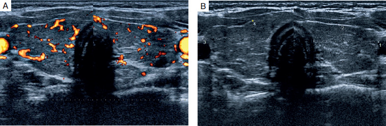
Thyroid ultrasonography revealed a pattern consistent with autoimmune thyroid disease (Figures 1A and 1B).
Figure 1
Ultrasonographic image of the patient’s thyroid at diagnosis – the gland is diffusely enlarged, heterogeneous, and deeply hypoechoic (A); power Doppler reveals mildly increased vascular flow (B)
In view of the clinical picture and laboratory results, Schmidt’s syndrome, one of the phenotypes of APS-2, was diagnosed. During hospitalisation, hydrocortisone was started intravenously at a total daily dose of 10 mg/m2 of body-surface area; after three days, levothyroxine 25 μg once daily was added, leading to a marked improvement in the patient’s general condition and well-being. Two weeks later, repeat clinical and biochemical evaluation revealed hyponatraemia (Na 130 mmol/l) and hyperkalaemia (K 5.19 mmol/l); treatment was therefore adjusted by adding fludrocortisone (50 μg in the morning and 25 μg in the evening). The girl’s clinical status improved, and the previously reported symptoms resolved.
Follow-up tests demonstrated normalisation of free thyroid hormone and cortisol concentrations, as well as of serum electrolytes. A decline in TSH and ACTH levels was also observed, confirming the efficacy of replacement therapy and restoration of both the hypothalamic–pituitary–adrenal and thyroid axes.
Discussion
A particularly important aspect of this case report is the occurrence of an adrenal crisis after levothyroxine was introduced before Addison’s disease had been diagnosed, underscoring the critical importance of the correct treatment sequence in APS-2. This patient’s course highlights the need for diagnostic vigilance in individuals with (very common) autoimmune hypothyroidism who present with nonspecific systemic symptoms. The sequential emergence of mutually masking manifestations and the clinical deterioration that follows inadequately managed hormone therapy should serve as a clinical alert, enabling the earliest possible recognition of the underlying disorder(s).
Schmidt’s syndrome, a form of APS-2, is relatively rare but potentially life-threatening, and its symptoms may develop slowly and non-specifically. APS-2 typically arises in young adults, more often in women, with a peak incidence in the second and third decades of life [3]. The complete triad is present in only about 10% of patients. The disease can evolve over many years, with the clinical features of the individual components appearing in sequence [4].
APS-2 results from dysfunction of the immune system, leading to the production of autoantibodies directed against endocrine-gland structures. Genetic factors play a significant role, particularly the presence of certain human leukocyte antigen (HLA) haplotypes, chiefly HLA-DR3 and HLA-DR4 [5]. Progressive destruction of glandular cells ultimately results in gland failure.
The clinical presentation of Schmidt’s syndrome is variable and depends on which component predominates and the degree of endocrine insufficiency. Most often the first manifestation is Addison’s disease, presenting with weakness, weight loss, hypotension, hypoglycaemia, hyponatraemia, hyperkalaemia, and characteristic skin hyperpigmentation [4]. In autoimmune thyroiditis there is a gradual onset of hypothyroidism with somnolence, cold intolerance, weight gain, constipation, and psychomotor slowing, symptoms that may be masked by coexisting adrenal insufficiency. Betterle et al. [5] pointed out that clinically overt disorders are only the tip of the iceberg of autoimmune disease, as latent forms are far more common. Nearly 70% of patients with Addison’s disease develop autoimmune thyroiditis, whereas the combination of adrenal insufficiency and type 1 diabetes occurs in approximately 52% of cases of Schmidt’s syndrome [6]. It is noteworthy that almost 40–50% of patients with Addison’s disease will, over the course of their lives, develop at least one additional autoimmune endocrine disorder. Accordingly, it is essential to carry out comprehensive screening for serum autoantibodies (such as anti-thyroid peroxidase, anti-thyroglobulin, anti-glutamic-acid-decarboxylase, and anti-islet-cell antibodies) [6].
The present case illustrates the classical evolution of Schmidt’s syndrome, with the sequential appearance of primary adrenal insufficiency followed by autoimmune hypothyroidism. Whenever one autoimmune disease is diagnosed (e.g. autoimmune thyroiditis), meticulous surveillance and evaluation for concomitant failure of other endocrine glands are crucial. Our patient’s pronounced skin hyperpigmentation could have been an important clue to primary adrenal insufficiency, although this sign tends to appear at a relatively late stage of the disease. Weight loss is likewise a significant feature because it is not seen in hypothyroidism. The patient’s complaints intensified after levothyroxine therapy had been initiated for hypothyroidism, most probably because levothyroxine enhances hepatic cortisol metabolism and increases cortisol clearance, thereby accelerating the onset of adrenal crisis in individuals with coexisting adrenal insufficiency [5, 7].
To establish the diagnosis of primary adrenal insufficiency, a cosyntropin (ACTH) stimulation test to assess cortisol secretory reserve is recommended, together with measurement of plasma ACTH, renin, and aldosterone. Treatment consists of replacement with both glucocorticoids and mineralocorticoids, i.e. hydrocortisone and fludrocortisone, respectively [8].
Clinical vigilance is paramount, particularly in patients who show an inadequate response to hormone therapy or clinical deterioration after levothyroxine initiation. Autoimmune thyroiditis is the most common endocrine, autoimmune, and thyroid disorder. In adrenal insufficiency, failure to institute appropriate therapy may be life-threatening, chiefly because of the risk of adrenal crisis; hence, accurate diagnosis and the sequential management of each hormonal disturbance in this syndrome are of critical importance.
Conclusions
The described case illustrates the diagnostic difficulties and the need for a comprehensive endocrine evaluation when systemic symptoms are present and therapy for a hormonally mediated disorder proves ineffective. In Schmidt’s syndrome, the correct treatment sequence is crucial, hydrocortisone replacement must be initiated first, followed by management of hypothyroidism, to prevent complications arising from severe, clinically overt hormonal decompensation in the course of Addison’s disease. Because of its nonspecific clinical picture, Schmidt’s syndrome is usually diagnosed late. Therefore, after any autoimmune disease is detected, screening for other potentially related endocrine pathologies is essential [3, 9, 10].