Przegląd Dermatologiczny
facebook
eISSN: 2084-9893
ISSN: 0033-2526
Dermatology Review/Przegląd Dermatologiczny
Bieżący numer Archiwum Artykuły zaakceptowane O czasopiśmie Zeszyty specjalne Rada naukowa Bazy indeksacyjne Prenumerata Kontakt Zasady publikacji prac Standardy etyczne i procedury
Panel Redakcyjny
Zgłaszanie i recenzowanie prac online
SCImago Journal & Country Rank
4/2025
vol. 112
 
Poleć ten artykuł:
Udostępnij:
Opis przypadku

Giant Keratoacanthoma Mimicking Squamous Cell Carcinoma in a Patient with Chronic Myeloid Leukemia

Beata Zagórska
1, 2
,
Karol Kołkowski
2, 3
,
Martyna Sławińska
1
,
Wojciech Biernat
4
,
Waldemar Placek
2

  1. Department of Dermatology, Venereology and Allergology, Medical University of Gdańsk, Poland
  2. Derm-Art, Training, Scientific and Research Center of Aesthetic Dermatology, Photodermatology and Dermatosurgery, Gdynia, Poland
  3. Department of Plastic Surgery, University Clinical Center, Gdańsk, Poland
  4. Department of Pathomorphology, Medical University of Gdańsk, Poland
Dermatol Rev/Przegl Dermatol 2025, 112, 249–252
Data publikacji online: 2025/10/30
Plik artykułu:
- Giant.pdf  [0.20 MB]
Pobierz cytowanie
 
Metryki PlumX:
 

INTRODUCTION

Keratoacanthoma (KA) is a tumor that arises from hair follicle epithelium. It is characterized by rapid growth, tendency to local invasion and spontaneous regression [1]. Typically, the tumor reaches its final size of 10 to 25 mm within 6 months [2]. Classical and most common KA variant manifests as a solitary, sharply demarcated, firm, pinkish or skin-colored nodule or tumor with a central hyperkeratotic horn plug, the removal of which creates a crater-like structure [3, 4]. Less common variants are keratoacanthoma centrifugum marginatum, giant keratoacanthoma (GKA), eruptive keratoacanthoma and multiple keratoacanthomas associated with genetic syndromes [5, 6]. GKA is a rare variant of solitary keratoacanthoma and is characterized by a size greater than 20 mm. It has been diagnosed more often in men with a peak incidence in the fifth decade of life [7]. In the differential diagnosis, mainly cutaneous squamous cell carcinoma (SCC) should be considered [4].

OBJECTIVE

This article presents the case of a patient with a giant keratoacanthoma in the context of chronic myeloid leukemia.

CASE REPORTS

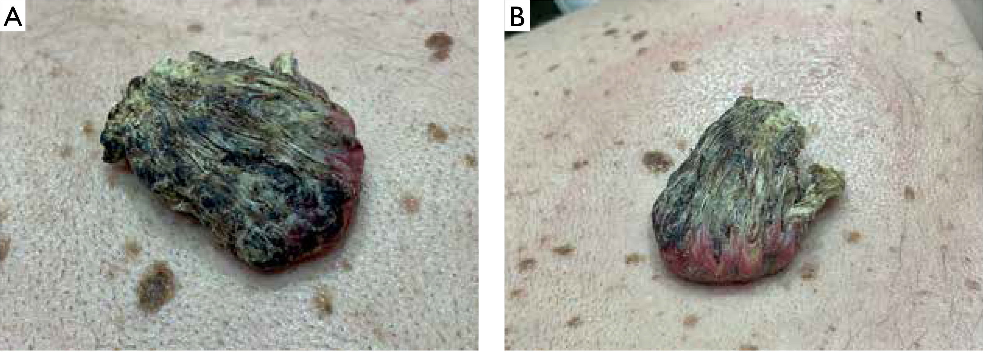
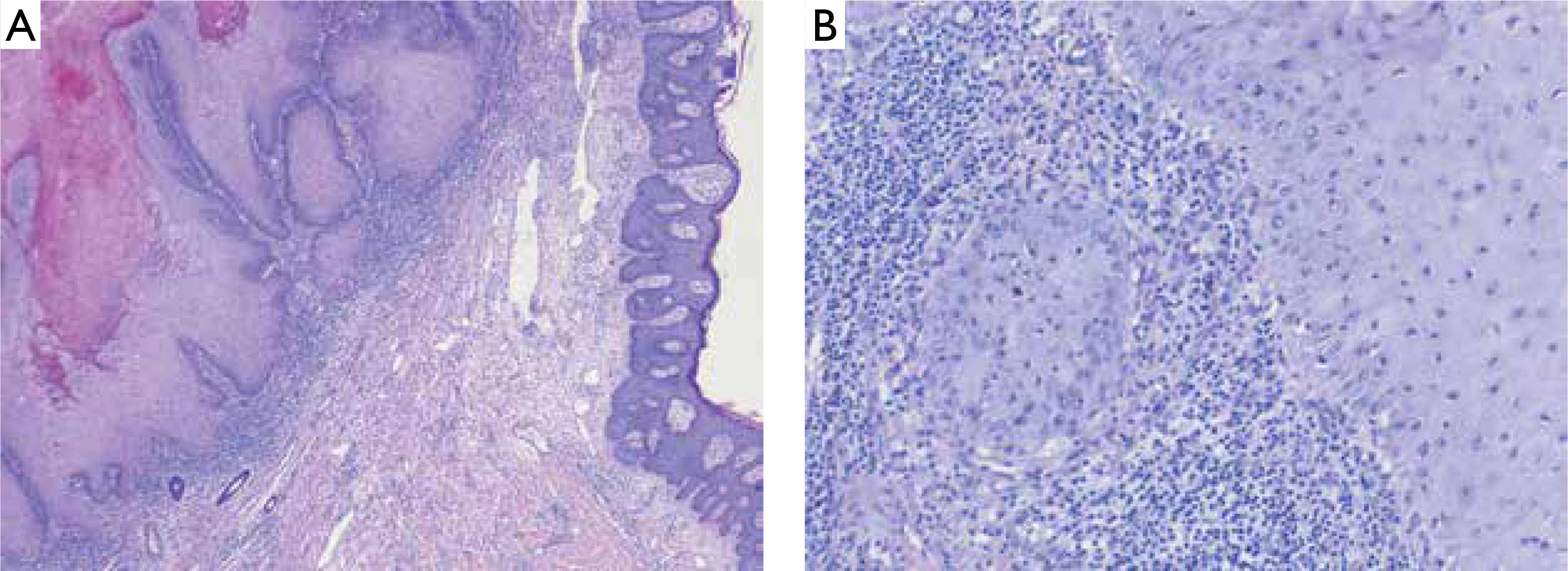
A 57-year-old man presented with a tender to palpation, large (10 × 6 cm in size) exophytic tumor with central hyperkeratotic masses located on his back, in the vertebral line of the interscapular region (figs. 1, A, B). The lesion had appeared approximately 2 years earlier and was growing with time. There was no history of preceding trauma. The patient suffered from chronic myeloid leukemia and schizophrenia, diagnosed over 25 years before. At the time of presentation, the patient was taking quetiapine and amisulpride. Besides, the clinical examination was insignificant. After obtaining the patient’s written consent, the lesion was surgically excised with 0.5 cm margin. Based on histopathological evaluation keratoacanthoma was diagnosed (fig. 2).

Figure 1

A, B – Clinical presentation. Sharply demarcated, pedunculated tumor measuring 10 × 6 cm with central hyperkeratotic masses

/f/fulltexts/PD/57020/PD-112-57020-g001_min.jpg
Figure 2

A, B – Histological presentation. A – Low magnification shows the marginal part of the tumor with a surrounding ‘collar’ of typical epidermis (right side). B – Lower part of the tumor with a ‘de-lacerating’ tumor nest surrounded by an intense infiltrate of lymphocytes

/f/fulltexts/PD/57020/PD-112-57020-g002_min.jpg

DISCUSSION

Giant KA is a rarely described tumor. According to the only report summarizing clinical and pathological features conducted by Bogner et al., GKA occurs predominantly on the face and limbs [8]. The largest GKA reported in a 55-year-old individual was located on the hand, developed within post-burn scar and measured 10 × 7 cm [1]. The characteristic feature of KA is the evolution of the tumor divided into three stages: rapid growth (4–14 weeks), stationary phase (2–8 weeks) and involution, usually lasting 4 months [913]. In the case of the described patient, the lesion gradually enlarged for 2 years and at the time of diagnosis did not show any signs of regression. Although a few similar cases of GKA with a longer history have been described, the exact evolution of giant tumors enabling clinical differentiation from SCC has not been known so far [8]. Similarly, the etiology and pathogenesis of the disease are debatable. The most common risk factors of KA include chronic exposure to ultraviolet (UV) and Roentgen radiation, contact with tar products, trauma, skin burns, human papillomavirus infection, certain medications (including PD-1/PD-L1, BRAF and Hedgehog pathway inhibitors, sorafenib, infliximab), as well as immunological disorders and post-infectious immunosuppression [1420]. Among the described cases of giant tumors, almost all were located on the skin chronically exposed to UV radiation and several of them were associated with pre-existing diseases such as burn or hyperplastic lichen planus [8]. History of chronic myeloid leukemia was the most important risk factor for GKA in our patient. The histopathological features of KA are marked acanthosis, hyperkeratosis and a central, rimmed (or supported) keratin plug at the edges giving a characteristic crater-like appearance [8, 21]. KA, including giant lesions, may microscopically resemble well-differentiated SCC, but the lack of vascular and neural invasion and preservation of the basement membrane integrity help to distinguish these two entities [21]. The most common treatment method reported in the literature is surgery and combined surgical treatment with radiotherapy [8, 22, 23]. Intralesional injections of cytostatic drugs have also been studied in several reports to accelerate regression of the lesion [23]. However, the efficacy of pharmacological treatment is difficult to assess due to the possibility of spontaneous tumor regression. Another limitation is that available data is based on case series with the absence of randomized controlled trials [22, 23]. Considering diagnostic difficulties in differentiation with SCC, high risk of post-involution scarring and lack of sufficient knowledge about the biological behavior of GKA, surgical excision seems to be the best treatment option.

CONCLUSIONS

Giant KA may be difficult to diagnose because of its atypical clinical presentation. The main differential diagnosis is SCC. Surgical excision with histopathological examination remains the standard therapeutic procedure.

ETHICAL APPROVAL

Not applicable.

CONFLICT OF INTEREST

The authors declare no conflict of interest.

References

1 

Wagner V.P., Martins M.D., Dillenburg C.S., Meurer L., Castilho R.M., Squarize C.H.: Histogenesis of keratoacanthoma: histochemical and immunohistochemical study. Oral Surg Oral Med Oral Pathol Oral Radiol 2015, 119, 310-317.

2 

Park H., Park H., Kim H., Yeo H.: A giant keratoacanthoma treated with surgical excision. Arch Craniofac Surg 2015, 16, 92-95.

3 

Nirenberg A., Steinman H., Dixon A.: Keratoacanthoma: update on the Debate. Am J Dermatopathol 2021, 43, 305-307.

4 

Sławińska M., Nowicki R., Sobjanek M.: Praktyczne aspekty diagnostyki i leczenia rogowiaka kolczystokomórkowego skóry. Dermatol Dypl 2020, 11, 15-19.

5 

Mehrtens S.H., de la Hera I., Shankar S.: Case of keratoacanthoma centrifugum marginatum treated with acitretin. BMJ Case Rep 2018, 2018, bcr2018226818.

6 

Vitiello M., Echeverria B., Romanelli P., Abuchar A., Kerdel F.: Multiple eruptive keratoacanthomas arising in a tattoo. J Clin Aesthet Dermatol 2010, 3, 54-55.

7 

Ni Y., Li R., Zhu D., Dong R., Qiao F.: Surgical excision of a giant solitary keratoacanthoma in the cheek: a case report and literature review. AME Case Rep 2023, 8, 7.

8 

Bogner P.N., Cheney R.T., Zeitouni N.C.: Giant keratoacanthoma: case report and review of the english literature. Am J Dermatopathol 2014, 36, 252-257.

9 

Lejkan K., Starzycki Z.: Giant keratocanthoma on the inner surface of the prepuce. Br J Vener Dis 1977, 53, 65-67.

10 

Kennedy D., Song R.: Growth on the abdomen. Am Fam Phys 2019, 99, 255-256.

11 

Pravdina O.V., Vodolazskij V.A., Naboka M.V., Ohlopkov V.A.: The case of recurrent aggregated giant keratoacanthoma with underlying post-burn scar. Clin Dermatol Venereol 2016, 15, 29-32.

12 

Chorzelski T.P., Jabłońska S., Błaszczyk M.: Keratoacanthoma multiplex – Varietas Grzybowski. Dalsza siedmioletnia obserwacja chorego w pełni potwierdza koncepcję autora. Przegl Dermatol 1995, 82, 405-411.

13 

Schwartz R.A.: Keratoacanthoma. J Am Acad Dermatol 1994, 30, 1-19.

14 

Letzel S., Drexler H.: Occupationally related tumors in tar refiner workers. J Am Acad Dermatol 1998, 39, 712-720.

15 

Tamir G., Morgenstern S., Ben-Amitay D., Okon E., Hauben D.J.: Synchronous appearance of keratoacanthomas in burn scar and skin graft donor site shortly after injury. J Am Acad Dermatol 1999, 40, 870-871.

16 

Norgauer J., Rohwedder A., Schaller J.: Human papillomavirus and Grzybowski’s generalized eruptive keratoacanthoma. J Am Acad Dermatol 2003, 49, 771-772.

17 

Mager L., Plaza J.A., Sopkovich J., Kaffenberger B.H., Dulmage B.: Eruptive keratoacanthoma secondary to immune checkpoint inhibitors: a narrative review. Arch Dermatol Res 2025, 317, 234.

18 

Jantzem H., Dupre-Goetghebeur D., Spindler P., Merrer J.: Kératoacanthomes multiples éruptifs induits par le sorafénib [Sorafenib-induced multiple eruptive keratoacanthomas]. Ann Dermatol Venereol 2009, 136, 894-897.

19 

Esser A.C., Abril A., Fayne S., Doyle J.A.: Acute development of multiple keratoacanthomas and squamosus cell carcinomas after treatment with infliximab. J Am Acad Dermatol 2004, 50, 75-77.

20 

Vanhooteghem O., Wiart T., Creusy C., de Beer P.: Multinodular keratoacanthoma and T cell lymphoma. J Eur Acad Dermatol Venerol 1996, 6, 50-56.

21 

Miedziński F.: Rogowiak kolczystokomórkowy w ujęciu patogenetycznym i klinicznohistologicznym, jego 40-letnia rocznica. Post Dermatol Alergol 1990, 7, 61-70.

22 

Collins A., Savas J., Doerfler L.: Nonsurgical treatments for nonmelanoma skin cancer. Dermatol Clin 2019, 37, 435-441.

23 

Seger E.W., Tarantino I.S., Neill B.C., Wang T.: Relative efficacy of nonoperative treatment of keratoacanthomas. J Cutan Med Surg 2020, 24, 41-46.

Copyright: © 2025 Polish Dermatological Association. This is an Open Access article distributed under the terms of the Creative Commons Attribution-NonCommercial-ShareAlike 4.0 International (CC BY-NC-SA 4.0) License (http://creativecommons.org/licenses/by-nc-sa/4.0/), allowing third parties to copy and redistribute the material in any medium or format and to remix, transform, and build upon the material, provided the original work is properly cited and states its license.
 
© 2025 Termedia Sp. z o.o.
Developed by Bentus.