
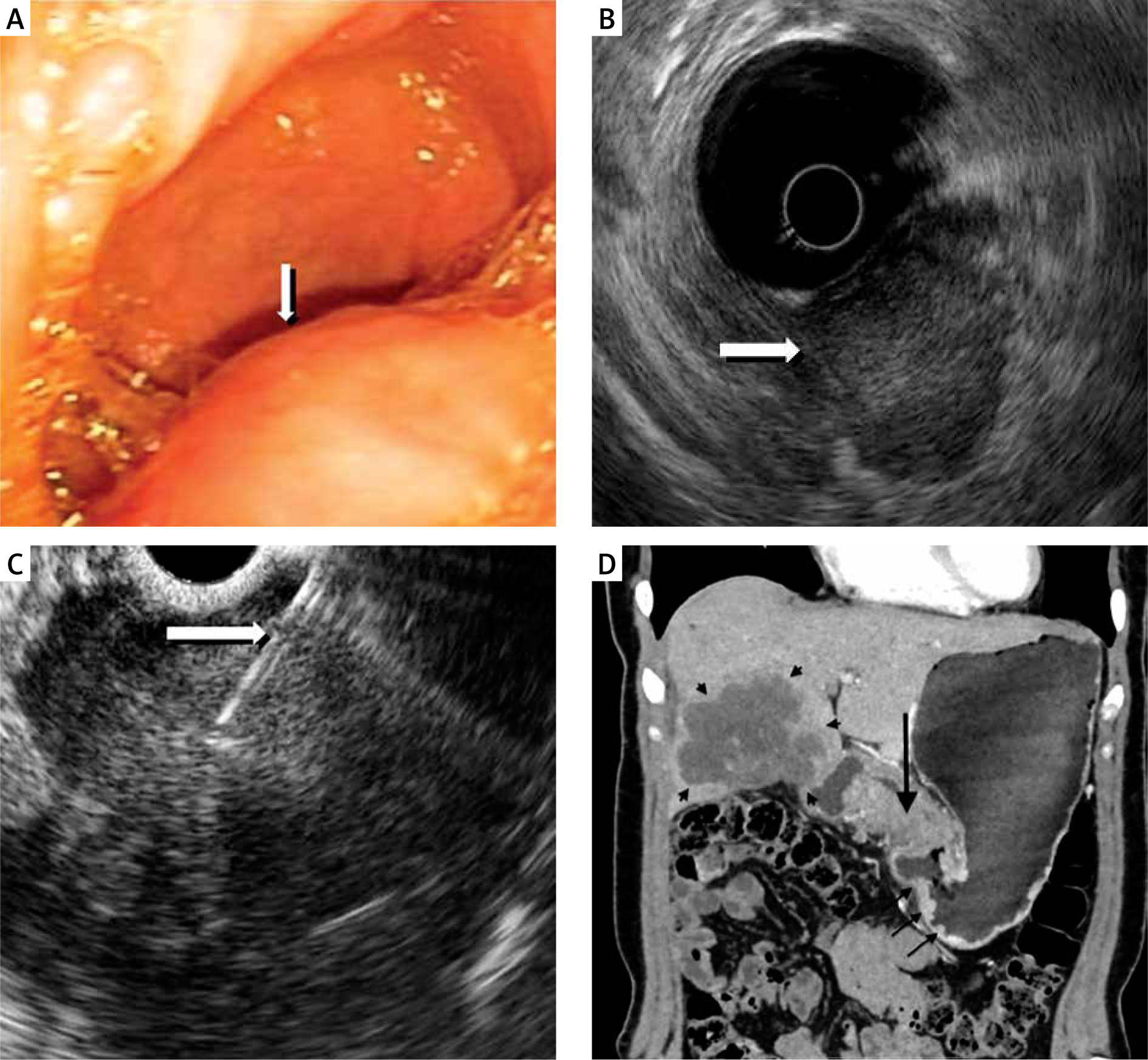
A 42-year-old woman was referred to the Gastroenterology Department because of colic, epigastric pain lasting over 3 months, with moderate improvement after analgesics/spasmolytics. She denied any other ailments. Abdominal ultrasound revealed a well-defined (42 × 32 mm) lesion, mostly solid with cystic components, near the gallbladder. Gastroscopy showed a subepithelial tumour at the posterior gastric wall of the prepyloric region, covered with normal mucosa (Figure 1 A).
Figure 1
A – A subepithelial tumour at the posterior gastric wall of the prepyloric region, covered with normal mucosa revealed in gastroscopy. B – Tumour, which invaded the muscularis propria and submucosa of gastric wall and presented mostly extraluminal growth. C – EUS-guided biopsy of the gastric mass. D – Contrast-enhanced CT (coronal MIP reconstruction) revealed a large heterogeneous mass replacing the gallbladder, with wide infiltration of adjacent structures (small arrows). Patchy contrast enhancement mass is seen in the antral part of the stomach (long arrow) with infiltration of the gastric wall (middle arrows)
Her past medical history was brief, with no chronic diseases, and included left-sided urolithotripsy, left inguinal hernia surgery, and 3 caesarean sections. Physical examination revealed only mild pain in the epigastrium at palpation. Routine laboratory tests revealed anaemia (Hgb = 9.7 g/dl, RBC 3.78 million/µl, HCT 30%) and high CRP (40.64 mg/dl). Other parameters (sodium and potassium levels, creatinine, urea, AST, ALT, GGTP, AP, bilirubin, CEA, AFP, and CA19-9) were normal.
The patient underwent endoscopic ultrasound (EUS), which showed a large subepithelial mass in the antral part of the stomach. The tumour invaded the muscularis propria and submucosa of the gastric wall and presented mostly extraluminal growth (Figure 1 B). It was suspected to be a metastatic lesion. At the porta hepatis, extensive heterogeneous infiltration with necrosis foci was visible. EUS-guided biopsy of the gastric mass was performed (Figure 1 C) and revealed keratinized squamous cell carcinoma.
A CT scan of the abdomen showed gallbladder cancer with metastatic spread: in the anatomical location of the gallbladder, a tumour 88 mm long, widely infiltrating segments 4a, 4b, and 5 of the liver; in the middle epigastrium, a solid mass 64 × 54 × 76 mm, infiltrating/compressing the antral part of the stomach, numerous metastases in the peritoneum, and enlarged lymph nodes (Figure 1 D). Chest X-ray/computed tomography (CT) were normal. No gynaecological abnormalities were found.
The patient was scheduled for chemotherapy with gemcitabine and carboplatin combo but did not show up for her first course of treatment. However, she is still alive 7 months after being diagnosed.
Gallbladder cancer (GBC) is a rare disease [1, 2], with adenocarcinoma as the most common (76–90% of cases) histological subtype. Squamous cell carcinoma (SCC) is very rare and represents approximately 2–10% of all cases [2]. According to PubMed there are only a few cases of this histological type of cancer. Also, the patient described is the first one with such a diagnosis in our multiannual clinical practice. SCC of the gallbladder is usually diagnosed at an advanced stage and has an extremely poor prognosis with an average overall survival time of 6 months. SCC has a worse prognosis than adenocarcinoma due to its more malignant and aggressive invasiveness to adjacent and distant structures, scarce early symptoms, and unresponsiveness to chemotherapy [2–4].
Gallbladder cancer can be associated with cholelithiasis and parasitic infections. Other risk factors include the following: obesity, smoking, diabetes, porcelain gallbladder, primary sclerosing cholangitis (PSC), and a diet rich in salted, preserved food [5].
Due to scarce clinical manifestation, diagnosis is mostly based on imaging techniques, such as abdominal ultrasonography, followed by CT or magnetic resonance imaging (MRI), which are essential. Definitive diagnosis needs histopathological expertise [1]. Surgery is the mainstay of treatment for patients with localized tumours. Unfortunately, most cases of GBC are inoperable [4]. Therefore, there is a need to search for new diagnostic methods at the early stage of the disease to enable surgical treatment and long-term survival.


